

Abstract
In this study, the effect of applying wharton jelly mesenchymal stromal cells (WJ-MSC) isolated from the human umbilical cord tissue on the neonatal mouse model caused experimental asphyxia in mice was investigated. WJ-MSC surface markers (CD44, CD90, CD105) were characterised by immunofluorescence staining, and pluripotency genes (Nanog, Oct-4, Sox-2) were characterised by qPCR. Blood, prefrontal cortex, cerebellum, hippocampus, lung, heart, kidney, and liver tissues were analysed twenty days after subcutaneously administered WJ-MSC. WJ-MSC administration significantly decreased serum TNF-α, NSE, GFAP, and IL-6 levels in the asphyxia mice. It was determined that WJ-MSC application in tissues accelerated cell regeneration and decreased oxidative stress. In conclusion, this study showed that multiorgan damage in asphyxia could be prevented by applying WJ-MSC at an early stage. Therefore, WJ-MSC application in infants with neonatal asphyxia in the clinic may be an innovative method in the future.
Graphical Abstract
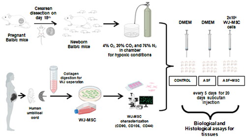
The umbilical cord was taken from human caesarean section and separated wharton jelly (WJ). WJ applied the enzymatic degradation by collagenase type 1. Mesenchymal stromal cells (MSC) isolated from WJ and characterised by CD90, CD105, and CD44 cell surface receptors staining. Newborn Balb/c mice taken by caesarean section on day 18th were placed in chambers and exposed to 4% O2, 20% CO2, and 76% N2 gas to hypoxic conditions for asphyxia. WJ-MSC (2 × 10⁶ cells) were administered subcutaneously to asphyxiated mice every five days for 20 days (n = 7). Multiple organ damage was performed on prefrontal cortex, cerebellum, hippocampus, heart, lung, kidney, and liver tissues using biological and histological assays.
What is already known on this subject? Perinatal asphyxia is a serious condition that generates progressive neurological defects and systemic multi-organ failure because of the absence of blood flow or gas exchange in the fetusfoetus during birth. There is no clinical application to reduce damage except for hypothermia treatment in asphyxia.
What do the results of this study add? This study showed that WJ-derived mesenchymal stromal cell administration could be used to treat hypoxia-induced asphyxia in newborns. In addition, it can be effective against multi-organ damage caused by asphyxia by evaluating the rich content of the UC tissue expelled during birth.
What are the implications of these findings for clinical practice and/or further research? In treating asphyxia, the need to develop new strategies emerges as these are not effective in reducing the damage adequately. This WJ-MSC will be new hope for treating asphyxia and guide clinical applications.
IMPACT STATEMENT
Acknowledgments
We thank ODC Research and Development Inc for their support in cell culture materials.