

Abstract
Objective
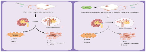
The purpose of this study was to determine the effect of tripterygium glycosides (TGs) on regulating abnormal lipid deposition in nephrotic syndrome (NS) rats.
Methods
Sprague-Dawley (SD) rats were injected with 6 mg/kg doxorubicin to construct nephrotic syndrome models (n = 6 per group), and then administered with TGs (10 mg/kg·d−1), prednisone (6.3 mg/kg·d−1), or pure water for 5 weeks. Biomedical indexes, such as urine protein/creatinine ratio (PCR), blood urea nitrogen (BUN), serum creatinine (Scr), serum albumin (SA), triglycerides (TG), total cholesterol (TC)were investigated to evaluate the renal injury of rats. H&E staining experiment was used to assess the pathological alterations. Oil Red O staining was used to assess the level of renal lipid deposition. Malondialdehyde (MDA) and glutathione (GSH) were measured to assess the extent of oxidative damage to the kidney. TUNEL staining was used to assess the status of apoptosis in the kidney. Western blot analysis was performed to examine the levels of relevant intracellular signaling molecules.
Results
After treatment with TGs, those tested biomedical indexes were significantly improved, and the extent of kidney tissue pathological changes and lipid deposition in the kidney was diminished. Treatment with TGs decreased renal oxidative damage and apoptosis. Regarding the molecular mechanism, TGs significantly increased the protein expression levels of Bcl-2 but decreased the levels of CD36, ADFP, Bax, and Cleaved caspase-3.
Conclusion
TGs alleviates renal injury and lipid deposition induced by doxorubicin, suggesting that it may be a new strategy for reducing renal lipotoxicity in NS.
Graphical Abstract
Ethical approval
The animal protocol was reviewed and approved by the Experimental Animal Ethics Committee of Guangdong Provincial Hospital of Traditional Chinese Medicine, approval number (2020004).
Author contributions
Bo Liu, Aihua Wu, and Peng Xu conceived and designed the experiments. Bidan Zheng, Dongfang Lu, Xiuping Chen, Yinghua Yin, and Weiying Chen performed the experiments. Bidan Zheng, Huanmei Lin, and Dongfang Lu analyzed and interpreted the data. Bidan Zheng and Dongfang Lu wrote the manuscript. Bo Liu and Aihua Wu critically revised the manuscript. Bo Liu, Aihua Wu, Peng Xu, and Xiaowan Wang supervised the findings of the work and approved the manuscript for submission. All authors agreed with the final version of this manuscript.
Disclosure statement
The graphical abstract was created by Figdraw. The authors declare no conflicts of interest.
Data availability statement
All data generated during the study can be obtained upon reasonable request from the corresponding author.