

Abstract
Objective: To evaluate the changes in cardiac morphology of fetuses with congenital heart disease (CHD) using the fetal heart quantitative technique (fetalHQ).
Methods: A total of 20 normal pregnant women (control group) and 20 pregnant women suspected of fetal CHD (case group) were included in this study. The dynamic images of the four-chamber view of the fetal heart were recorded and analyzed using fetalHQ. The global sphericity index (GSI) and 24-segment SI of the two groups were compared. The differences in the left and right ventricular 24-segment SI for each group were investigated.
Results: There was no statistically significant difference in the GSI between the two groups (p > 0.05). The difference in the SI values of left ventricular segments 1–2 between the case group and control group was statistically significant (all p < 0.05), while the intergroup difference in SI of left ventricular segments 3–24 was not significant (all p > 0.05). The SI of the 24 segments of the right ventricle showed no significant intergroup difference (all p > 0.05). The difference in the left and right ventricular 24-segment SI in the case group did not reach statistical significance (all p > 0.05). In the control group, the SI values between the left and right ventricles were significantly different in segments 18–24 (all p < 0.05), and no significant difference was found in segments 1–17 (all p > 0.05). There was a statistically significant intergroup difference in the percentage of unusual left ventricular SI, determined based on Z-score (p < 0.05), and the percentage of outliers for the right ventricle between the two groups showed no significant difference (p > 0.05).
Conclusion: The fetalHQ is regarded as a straightforward and reliable approach for assessing the cardiac GSI and 24-segment SI of left and right ventricles in fetuses diagnosed with CHD. While CHD may not significantly impact the overall shape of the fetal heart or the geometric shape of the right ventricle, in this study, a notable increase in SI values for the left ventricular 1–2 segments was observed, indicating a more flattened ventricular chamber. Additionally, the morphological distinctions between the left and right ventricles in fetuses with CHD are no longer discernible.
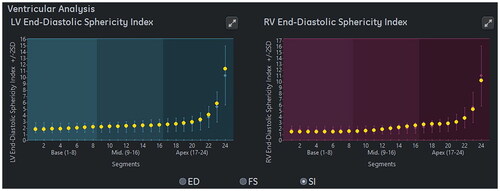
Graphical Abstract
Disclosure statement
The authors declare no potential conflicts of interest with respect to the research, authorship, and/or publication of this article.
Data availability statement
The data are available from the corresponding author on reasonable request.