Abstract
Coronary angiography (Figure‐) and computerized tomographic angiography (CTA) (Figure‐) of a 61‐year‐old man with exertional angina demonstrated proximal compression by the great vessels of an anomalous right coronary arising from the left main trunk. A significant left anterior descending lesion was also present. The patient underwent uneventful bypass surgery.
CTA is very useful for confirmation of the inter‐arterial course of an aberrant coronary artery. Proposed mechanisms of sudden death include exertional coronary compression between the great vessels, with associated ostial narrowing, as well as acute angulation of the coronary course. Coronary bypass is the treatment of choice for these patients. Citation
Figure 1. (a) Right‐anterior‐oblique coronary angiography of RCA, demonstrating a ‘slit‐like’ ostium. (b) Left‐anterior‐oblique coronary angiography of RCA, revealing common origin with left main trunk (arrow).
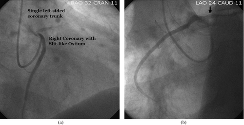
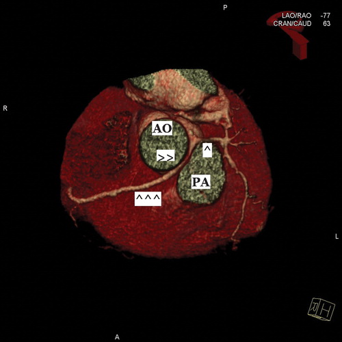
Figure 2. Three‐dimensional reconstruction of the heart by CTA demonstrating common single left‐sided coronary ostium giving rise to the left main coronary (single arrow) and proximal right coronary (double arrows) segments. Note the inter‐arterial course of the latter between the aorta (Ao) and pulmonary artery (PA), as well as the relatively narrowed diameter of this segment when compared to the normal‐caliber mid vessel (triple arrows).