

ABSTRACT
Vaccines utilizing modified messenger RNA (mRNA) technology have shown robust protective efficacy against SARS-CoV-2 in humans. As the virus continues to evolve in both human and non-human hosts, risk remains that the performance of the vaccines can be compromised by new variants with strong immune escape abilities. Here we present preclinical characterizations of a novel bivalent mRNA vaccine RQ3025 for its safety and effectiveness in animal models. The mRNA sequence of the vaccine is designed to incorporate common mutations on the SARS-CoV-2 spike protein that have been discovered along the evolutionary paths of different variants. Broad-spectrum, high-titer neutralizing antibodies against multiple variants were induced in mice (BALB/c and K18-hACE2), hamsters and rats upon injections of RQ3025, demonstrating advantages over the monovalent mRNA vaccines. Effectiveness in protection against several newly emerged variants is also evident in RQ3025-vaccinated rats. Analysis of splenocytes derived cytokines in BALB/c mice suggested that a Th1-biased cellular immune response was induced by RQ3025. Histological analysis of multiple organs in rats following injection of a high dose of RQ3025 showed no evidence of pathological changes. This study proves the safety and effectiveness of RQ3025 as a broad-spectrum vaccine against SARS-CoV-2 variants in animal models and lays the foundation for its potential clinical application in the future.
Data availability
Data supporting the findings in this study are included in the main article and associated files.
Results
Antigen design and characterization
RQ3025 is a bivalent broad-spectrum mRNA vaccine comprising two distinct mRNA molecules, namely RQ3013 and RQ3019, both based on the S protein sequences of SARS-CoV-2 variants. Both mRNAs encode a nearly full-length S protein excluding the eighteen cysteine-rich residues at the C-terminus. The S protein encoded by RQ3013 contains all mutations present in the B.1.1.7 variant, along with additional K417N, E484 K, and A701 V mutations found in the B.1.351 variant. Seven of these mutations also exist in the Omicron variant B.1.1.529 ((A)). RQ3019 incorporates all mutations from the BA.2 variant, with additional L452R and F486 V mutations found in the BA.4/BA.5 variant ((A)). Unlike the S2P design employed in BNT162b2 and mRNA-1273 vaccines [Citation17,Citation18], Immunogens encoded by RQ3013 and RQ3019 do not contain the two proline substitutions. Instead, the furin cleavage site 682RRAR685 is replaced by GSAS to prevent protease-mediated proteolysis. The native signal peptide and transmembrane domain are retained in both S proteins.
Figure 1. Vaccine design of RQ3025 and structural characterization of the expressed antigens. (A) An illustration of the composition of the RQ3025 Bivalent Vaccine, containing two mRNA sequences (RQ3013 and RQ3019) encoding the Spike protein of SARS-CoV-2. The furin cleavage site RRAR was replaced by GSAS.The last 18 amino acids were deleted. (B) Heatmap analysis of the mutation sites of the bivalent mRNA vaccine RQ3025 and the mutation sites of prevalent variant strains. (C) Western blots using different spike protein antibodies showing specificities of the antigens expressed by HEK293 T cells transfected with RQ3013, RQ3019, RQ3025, and Control. Purified RQ3013 and RQ3019 proteins were run in parallel on the same gels as positive control. (D) Localizations of the RQ3025, RQ3013 and RQ3019 RNA-encoded Spikes in HEK293 T cells transfected with the vaccine constructs were determined by immunofluorescence. Scale bar, 100 μm.
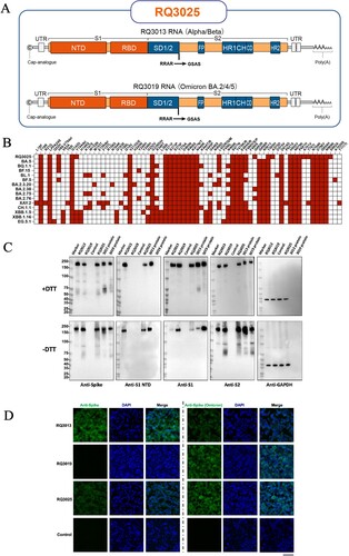
The mutation sites in RQ3025 cover all critical mutations found in 14 different variant strains including current VOIs such as BA.2.75, XBB.1.5, and VUMs such as XBB.1.16 and CH.1.1 ((B)). Well-established lipid nanoparticles (LNPs) are used for mRNA encapsulation. The mRNA encapsulation efficiency (EE) for all three LNP vaccine candidates exceeded 90%, with an average diameter of 70 nm (Figure S1A). Moreover, all LNPs exhibited a consistent polydispersity index (PDI) of approximately 0.1, indicative of a uniform particle size distribution (Figure S1A). Furthermore, LNPs encapsulating mRNA consistently exhibited a negative surface charge (Figure S1B). The immunoreactivity of antigens derived from RQ3025 was assessed using multiple antibodies targeting distinct domains of the SARS-CoV-2 spike protein in western blots. The antibodies exhibited discriminative recognition of S proteins derived from RQ3013 or RQ3019 transfected HEK293 T cells on western blots, demonstrating mutation-directed immune specificity of the S proteins ((C)). Cell lysates from HEK293 T transfected with RQ3013, RQ3019, or RQ3025 displayed bands similar to the purified proteins derived from RQ3013 and RQ3019 in the blots. These bands were consistent across all tested antibodies and conditions, with or without DTT. This confirms the integrity of the translated antigens from the mRNAs ((C)). Cellular localization of the translated antigens from RQ3025 mRNA was assessed by immunofluorescence using antibodies recognizing either the receptor binding domain (RBD) of the spike protein from SARS ((D), left panel) or SARS-CoV-2 (Omicron)-RBD ((D), right panel). The SARS-RBD antibody exclusively detected signals on the surface of RQ3013 transfected cells. In contrast, the SARS-CoV-2 (Omicron)-RBD antibody detected signals in both RQ3013 and RQ3019 transfected cells. This difference reflects the antibody evasion ability of the Omicron variants. Both antibodies displayed signals localized to the membrane in RQ3025-transfected cells ((D)). These results demonstrate the correct expressions and the immune characterizations of the antigens derived from RQ3025 in vitro.
Immunogenicity of RQ3025 in BALB/c mice
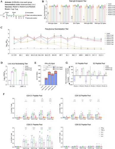
To assess the immunogenicity of RQ3025 in BALB/c mice, two groups (n = 24) were immunized intramuscularly with either 2 μg or 5 μg of RQ3013, RQ3019, or RQ3025 on day 0. Control mice (n = 24) received saline ((A)). All groups received booster injections on day 21. No local inflammation or other adverse effects were observed throughout the experiment. Sera were collected on day 28 to measure the immunobinding of IgGs to RBD, S and S2 by enzyme-linked immunosorbent assay (ELISA). RQ3013, RQ3019, and RQ3025 induced equally high titers of IgG binding to RBD, S, and S2 from the wild-type, B.1.351, and BA.5 variants ((B)). There was no difference between the low-dose (2 μg) and the high-dose (5 μg) groups immunized with the same vaccine ((B)). Next, we performed the lentiviral pseudovirus neutralization assay to evaluate the levels of neutralizing antibodies induced by RQ3013, RQ3019 or RQ3025 on day 28. RQ3013 elicited high titers of neutralizing antibodies against the B.1.351 variant in BALB/c mice. For several Omicron subvariants, such as BA.1, BA.2, BA.2.3.20, BA.2.38, BA.2.75, and BA.4/BA.5, RQ3013 also induced an effective amount of neutralizing antibodies. However, the neutralizing capacity of RQ3013 against BF.7, BL.1, BQ.1.1, XBB.1, XBB.1.5, CH.1.1 and EG.5.1 subvariants was significantly reduced ((C)). In contrast, RQ3019 induced high titers of neutralizing antibodies against various Omicron subvariants, albeit with subdued neutralization ability against B.1.351 ((C)). Immunization with the bivalent vaccine RQ3025 in BALB/c mice generated high titers of neutralizing antibodies against B.1.351 and all tested Omicron subvariants, e.g. geometric mean titer (GMT) = 4846 for XBB.1.5 and GMT = 1918 for EG.5.1 ((C)). Sera were collected from the RQ3025-immunized mice on day 42 for assessing the neutralizing titer against two live virus strains (B.1.351 and XBB.1.5) by Focus Forming Assay (FFA). In mice that received a 2-µg dose, the FRNT50 GMT of neutralizing antibodies against B.1.351 and XBB.1.5 were 3286 and 1064, respectively. A slightly higher neutralizing titer against B.1.351, but not XBB.1.5, was observed in mice that received a 5-µg dose ((D)). These results demonstrate excellent immunogenicity and broad neutralizing capacity of the bivalent mRNA vaccine RQ3025. Furthermore, long-term monitoring of the neutralizing antibody levels targeting B.1.351 and BA.4/BA.5 in BALB/c mice immunized with RQ3025 revealed persistent high titers of neutralizing antibodies against B.1.351 and BA.4/BA.5 in the mice 24 weeks post-immunization (Figure S2). Cell-mediated immunity was evaluated in the BALB/c mice immunized with RQ3025 or RQ3013 by using an IFN-γ ELISpot assay on isolated spleen cells restimulated with a pool of peptides derived from the S1 or S2 subunit. Both vaccines at low and high doses led to a strong cellular immune response in the spleen 3 months after the last immunization ((E)). Intracellular cytokine staining assays measuring expressions of IFN-γ, TNF-α, IL-2, IL-4 or IL-5 in S1 or S2 peptides stimulated spleen cells showed polyfunctional CD4+ and CD8+ T cell responses were induced by RQ3025 immunization, with a higher CD8+ T cell response for S1 than S2 ((F)). The T cell response induced by RQ3025 was comparable to that of RQ3013, indicating that the difference between RQ3025 and RQ3013 had little impact on cellular immunity ((E,F)). It has been reported that vaccine-associated enhanced respiratory disease (VAERD) was associated with a Th2-dominant immune response in SARS-CoV-2 infected hamsters [Citation19]. Therefore, the cytokines secreted by spleen cells upon restimulation with S1 and S2 peptide pools were evaluated to determine the T cell response type in RQ3025 immunized mice. The ratio of IFN-γ (Th1-associated cytokine) concentration to IL-4, IL-5, and IL-10 (Th2-associated cytokines) was 10–100 in S1 peptides stimulated splenocytes and approximately 10 in S2 peptides stimulated splenocytes, suggesting RQ3025 induced a Th1-dominant cell response ((G)).
Figure 2. Immunogenicity of RQ3025 in BALB/c mice. (A) The immunization scheme for RQ3013, RQ3019 and RQ3025 in mice. Mice (n = 24 per group) were intramuscularly immunized with saline (Control), low dose (2 μg) or high dose (5 μg) of RQ3013, RQ3019 or RQ3025. Time points of vaccination, bleeding, and splenocyte isolation are indicated by arrows. Eight or six mice from each group were randomly selected for ELISA or neutralizing activity measurement, respectively, while the rest were used to detect the T cell responses. (B) IgG antibody responses were measured by analyzing the binding of antibodies to the wild-type RBD antigen, S proteins from the wild-type and SARS-CoV-2 variants, and the wild-type S2 subunit using sera collected on day 28 via enzyme-linked immunosorbent assay (ELISA). Values are geometric mean ± SD. (C) Neutralizing antibody titers in sera collected on day 28, analyzed by the lentiviral luciferase-based pseudovirus assay. The black dashed line indicates the assay's detection limit (reciprocal titer of 12.5). Values are geometric mean ± SD. (D) The FFA-based live virus micro-neutralization assay of week-6 sera against SARS-CoV-2 B.1.351 and XBB.1.5. The black dashed line indicates the assay's detection limit (reciprocal titer of 16). Values are geometric mean ± SD. (E) Summed IFN-γ ELISpot responses in splenocytes restimulated with peptides spanning the entire S protein. The splenocytes were collected on day 113. SFU, spot-forming units. Data are presented as mean ± SEM. (F) CD4+ and CD8+ T cell responses in isolated splenocytes at 92 days post-boost stimulated with either vehicle or pools of overlapping peptides derived from the SARS-CoV-2 S protein for 5 h, shown by flow cytometry analyses of IFN-γ, IL-2, TNF-α, IL-4 and IL-5 expressions. Background cytokine expression in the presence of vehicle alone was accounted for and subtracted from the measured responses elicited by the S1 and S2 peptide pools for each individual mouse. Data are presented as mean ± SEM. The significance analysis was conducted in comparison with the control group. (G) MSD assay showing IFN-γ, IL-4, IL-5, and IL-10 cytokine levels secreted by T cells restimulated with S1 and S2 peptide pools in isolated splenocytes 46 days post-boost. Data are presented as mean ± SEM. Statistical analyses were carried out by ANOVA and Tukey’s multiple comparison tests (*P < 0.05; **P < 0.005; ***P < 0.001; ****P < 0.0001).
Immunogenicity of RQ3025 in K18-hACE2 transgenic mice
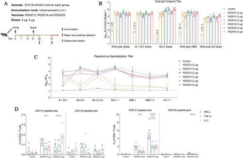
We next assessed the immunogenicity and efficacy of RQ3013, RQ3019 and RQ3025 in the K18-hACE2 transgenic mouse strain which is susceptible to SARS-CoV-2 and suffers from lung injuries that mimic severe SARS-CoV-2 infection in humans [Citation20–22]. Each mouse received two doses of either 2 or 5 μg injected on day 0 and day 21 ((A)). Both the 2- and 5-μg doses of all three vaccines elicited high titers of RBD- and S-specific IgG antibodies in the sera of K18-hACE2 mice at 7 days post-boost ((B)). Neutralizing antibodies in the sera were assessed by cell-based pseudovirus entry assay using viruses pseudotyped with the spike proteins of variant strains B.1.351, BA.4/5, BA.2.3.20, BQ.1.1, XBB.1, XBB.1.5, and CH.1.1. In consistence with the observations in BALB/c mice, RQ3025 elicited high levels of neutralizing antibodies against both the B.1.351 variant and the Omicron subvariants in comparison with RQ3013 and RQ3019 ((C)). Administration of two doses of RQ3025 (2 µg) induced neutralizing antibody titers of GMT 1878, 4246, 2792, 18,905, 2984, 681 and 1740 against B.1.351, BA.4/BA.5, BA.2.3.20, BQ.1.1, XBB.1, XBB.1.5, and CH.1.1, respectively ((C)). Spleen cells were isolated from the K18-hACE2 mice immunized with RQ3025 two months after the second injection and stimulated with a pool of S1 or S2 peptides. Cell-mediated immune response was triggered, which was evident from the elevated percentages of IFN-γ, TNF-α and IL-2 expressing CD4+ and CD8+ T cells ((D)). To assess the durability of immunity, serum samples were collected at 40 weeks post-initial immunization, and the presence of neutralizing antibodies was determined via a pseudovirus entry assay. The data revealed a gradual decline in the levels of neutralizing antibodies against both the B.1.351 variant and the Omicron subvariants from 4 weeks to 36 weeks, with these levels stabilizing by the 40th week (Figure S3A). In terms of the cellular immune response, splenocytes were harvested 40 weeks after the second immunization and subsequently stimulated with the same pool of peptides. A consistent immune response was observed when compared to the response at 2 months (Figure S3B).
Figure 3. Immunogenicity of RQ3025 in K18-hACE2 transgenic mice. (A) The scheme of mice immunization. Mice were intramuscularly immunized with saline (Control), low dose (2 μg) or high dose (5 μg) of RQ3013, RQ3019 or RQ3025 (n = 16 per group). Time points of vaccination, bleeding, and splenocyte isolation are indicated by arrows. (B) ELISA analysis of IgGs binding to the wild-type RBD antigen, S proteins from the wild-type and SARS-CoV-2 variants, and the wild-type S2 subunit with sera collected on day 28. Values are geometric mean ± SD. (C) Neutralizing antibody titers in day-28 sera, analyzed by the lentiviral luciferase-based pseudovirus entry assay. The black dashed line indicates the detection limit of the assay (reciprocal titer of 12.5). Values are geometric mean ± SD. (D) The percentages of CD4+ and CD8+ T cells expressing IFN-γ, IL-2, and TNF-α were assessed using intracellular cytokine staining and flow cytometry in splenocytes collected 60 days after the second immunization and stimulated with a pool of peptides derived from either the S1 or S2 regions of the SARS-CoV-2 Spike protein. Data are presented as mean ± SEM. The significance analysis was conducted in comparison with the control group. Statistical analyses were carried out by ANOVA and Tukey’s multiple comparison tests (**P < 0.005; ***P < 0.001; ****P < 0.0001).
Protection against SARS-CoV-2 variants in RQ3025 vaccinated K18-hACE2 transgenic mice
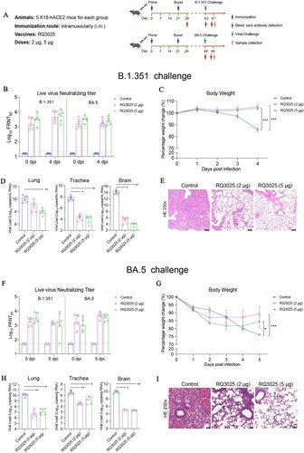
To measure the protective effect of RQ3025, K18-hACE2 mice vaccinated with RQ3025 or saline were challenged intranasally with B.1.351 (1 × 10³ PFU) 42 days post-boost or BA.5 (0.7 × 10⁵ PFU) 23 days post-boost ((A)). High live virus neutralizing titers against both B.1.351 and BA.5 variants were detected at 0 and 4 days post-infection (4 dpi) in sera from mice vaccinated with RQ3025. In addition, neutralizing antibody titers against B.1.351 in RQ3025-vaccinated mice were higher at 5 dpi than at 0 dpi ((B)). Mice infected with B.1.351 suffered more than a 10% body weight loss from 2 to 4 dpi, whereas those vaccinated with RQ3025 maintained their body weights ((C)). An significant reduction in viral load in the lung, trachea, and brain was detected in RQ3025-vaccinated mice at 4 dpi ((D)). Lung tissues at 4 dpi were sectioned and examined by hematoxylin and eosin (H&E) staining to evaluate lung injuries induced by B.1.351 infection. Control mice infected with B.1.351 exhibited severe lung injuries as evidenced by alveolar collapse, thickened alveolar septa, and extensive infiltration of inflammatory cells at 4 dpi, whereas mice vaccinated with RQ3025 showed mild to moderate inflammatory cell infiltration, and alveolar structures were maintained ((E) and Figure S5A). In mice challenged with BA.5 infection high titers of live virus neutralizing antibodies against both B.1.351 and BA.5 variants were also detected at 0 and 5 dpi in mice vaccinated with RQ3025 and neutralizing antibody titers against BA.5 in RQ3025-vaccinated mice were higher at 5 dpi than at 0 dpi ((F)). Control mice infected with BA.5 experienced over a 20% body weight loss from 1 to 5 dpi, whereas in mice vaccinated with RQ3025 (2 and 5 µg) body weight loss was averted from 3 dpi ((G)). RQ3025 vaccination significantly reduced the viral load in the lung, trachea and brain at 5 dpi ((H)). Lung tissues in RQ3025-vaccinated mice were protected from BA.5-induced injuries ((I) and Figure S5B). Together, RQ3025 induced humoral and cellular immune responses, providing protection against B.1.351 and BA.5 infection in K18-hACE2 mice.
Figure 4. Protective efficacy of RQ3025 in K18-hACE2 transgenic mice. (A) The scheme of mice immunization. Mice were intramuscularly immunized with saline (Control) or low dose (2 μg) or high dose (5 μg) of RQ3025 (n = 5 per group). Time points of vaccination, viral challenge, and sample collection are indicated by arrows. Data collected from mice challenged with B.1.351 are shown in (B-E). (B) Live virus neutralization titers at 0 dpi and 4 dpi were detected by the FFA-based live virus microneutralization assay. The black dashed line indicates the detection limit of the assay (reciprocal titer of 16). Values are geometric mean ± SD. (C) Monitoring of body weight changes in K18-hACE2 mice following infection with the variant B.1.351. Data are presented as mean ± SEM. (D) Viral load in the lungs, tracheae and brains at 5 dpi was determined by RT-qPCR. Data are presented as geometric mean ± SD. (E) Histopathological examinations (hematoxylin and eosin (H&E) staining) of lungs from the challenged mice at 5 dpi. Scale bar, 100 μm. Data collected from mice challenged with BA.5 are shown in (F-I). (F) Live virus neutralization titers at 0 and 5 dpi were detected by the PRNT-based neutralization assay. The black dashed line indicates the detection limit of the assay (reciprocal titer of 50). Values are geometric mean ± SD. (G) Monitoring of body weight changes in K18-hACE2 mice following infection with the variant BA.5. Data are presented as mean ± SEM. (H) Viral load in the lung, trachea and brain tissues at 5 dpi was determined by RT-qPCR. Data are presented as geometric mean ± SD. (I) Histopathological examinations (H&E staining) of the lungs from infected mice at 5 dpi. Scale bar, 100 μm. Statistical analyses were carried out by ANOVA and Tukey’s multiple comparison tests (*P < 0.05; **P < 0.005; ***P < 0.001).
Immunogenicity and protection of RQ3025 in hamsters
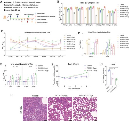
The Syrian hamster, expressing the ACE2 receptor, is highly susceptible to SARS-CoV-2 and develops pneumonia similar to that in COVID-19 patients, making it a suitable model for vaccine evaluation [Citation23]. Seven groups of hamsters were vaccinated on day 0 and day 21 with saline or the three vaccines (RQ3013, RQ3019 and RQ3025) at two doses (5 or 25 μg) followed by immunogenicity evaluation and viral challenge ((A)). High titers of antibodies binding to the RBD, S (B.1.351 and BA.5) and S2 subunit were detected in sera collected from RQ3025-immunized hamsters on day 28 ((B)). A pseudovirus neutralization assay with a panel of variant strains was used to test the range of neutralizing antibodies elicited by the three vaccines. RQ3025 elicited neutralizing antibodies against all variants while neutralizing antibodies induced by RQ3013 and RQ3019 were effective only against either B.1.351 or Omicron subvariants ((C)). Immunization with RQ3025 at a 25 μg dose elicited serum neutralizing antibody titers averaging 3246, 2941, 900, 2181, and 1120 against B.1.351, BA.4/BA.5, BA.2.3.20, BQ.1.1, and XBB.1 strains, respectively ((C)). Neutralizing titers against live virus B.1.351 and BA.5 were measured using a live virus microneutralization assay which also demonstrated the effectiveness of RQ3025 against both strains in comparison to the limited neutralizing abilities of RQ3013 and RQ3019 ((D)). Twenty-two days following the second vaccination, hamsters were challenged with the BA.5 variant (0.7 × 10⁵ PFU). Neutralizing antibodies against both B.1.351 and BA.5 live viruses in sera were detected at 0 and 5 dpi. A significant increase in neutralizing antibody against BA.5 was induced at 5 dpi and was further enhanced in hamsters vaccinated with 25 µg RQ3025 ((E)). Body weights of control hamsters slightly decreased at 5 dpi, whereas hamsters vaccinated with 25 µg RQ3025 exhibited an increase in body weight from 3 to 5 dpi. The 5-µg dose of RQ3025 immunization did not show an effect on body weight compared to the control ((F)). The viral load in the lungs of hamsters immunized with both doses of RQ3025 was significantly lower than that in the control at 5 dpi ((G)). Hamsters in the control group suffered lung injuries with thickened alveolar septa and a large number of inflammatory cells infiltration at 5 dpi while hamsters immunized with RQ3025 (5 and 25 µg) showed mild to moderate inflammatory cell infiltration and mild alveolar epithelial hyperplasia ((H) and Figure S5C). Together, these results provided evidence that RQ3025 vaccination elicits protection against SARS-CoV-2 variants in Syrian hamster.
Figure 5. Immunogenicity and protective efficacy of RQ3025 in hamsters. (A) The immunization scheme for hamsters by RQ3013, RQ3019 and RQ3025. Hamsters were intramuscularly immunized with saline (Control), low dose (5 μg) or high dose (25 μg) of RQ3013, RQ3019 or RQ3025 (n = 10 per group). Time points of vaccination, bleeding, viral challenge, and sample collection are indicated by arrows. (B) Binding of IgGs to the wild-type RBD, S proteins from the wild-type and SARS-CoV-2 variants and the wild-type S2 subunit analyzed with sera collected on day 28 by ELISA. Values are geometric mean ± SD. (C) Neutralizing antibody titers in day-28 sera analyzed by the lentiviral luciferase-based pseudovirus assay. The black dashed line indicates the assay's limit of detection (reciprocal titer of 12.5). Values are geometric mean ± SD. (D) The FFA-based live virus microneutralization assay of week-6 sera against SARS-CoV-2 B.1.351 and BA.5. The black dashed line indicates the assay's detection limit (reciprocal titer of 16). Values are geometric mean ± SD. (E) Live virus neutralization titers at 0 and 5 dpi were detected by the PRNT-based neutralization assay. The black dashed line indicates the detection limit of the assay (reciprocal titer of 50). Values are geometric mean ± SD. (F) Body weight changes of hamsters following infection with the variant BA.5. Data are presented as mean ± SEM. (G) Viral load in the lungs at 5 dpi was determined by RT-qPCR. Data are presented as geometric mean ± SD. (H) Histopathological examinations (H&E staining) of the lungs from infected hamsters at 5 dpi. Black scale bar, 100 μm. Statistical analyses were carried out by ANOVA and Tukey’s multiple comparison tests (**P < 0.005; ****P < 0.0001).
Immunogenicity of RQ3025 as a third booster in BALB/c mice
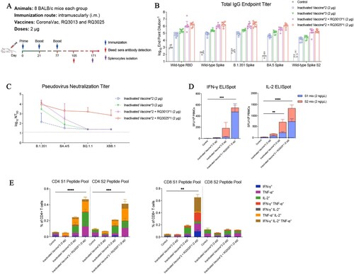
We next investigated the feasibility of mRNA vaccines as a booster following inoculation with inactivated vaccines. Mice immunized with two doses of inactivated vaccines on day 0 and day 21 received a third booster with an inactivated vaccine or mRNA vaccines RQ3013 or RQ3025 on day 77 (56 days after the second dose of the inactivated vaccine) ((A)). Sera were collected on day 105 for assessment of antibodies. A booster with either RQ3013 or RQ3025 elicited higher titers of RBD-, S-, and S2-specific IgGs than a booster with the inactivated vaccine ((B)). A booster with RQ3025 was the most effective in inducing specific antibodies binding to the BA.5 S protein ((B)). RQ3025 effectively boosted the neutralizing antibodies against both B.1.351 and Omicron variants (BA.4/BA.5, BQ.1.1 and XBB.1) whereas the third boost with the inactivated vaccine or RQ3013 failed to enhance the neutralizing capability against BQ.1.1 and XBB.1 ((C)). Splenocytes were isolated on day 171 (3 months after the third boost) for the assessment of cell-mediated immunity. RQ3025 induced higher cellular immune response than the inactivated vaccine, as measured by the amount of IFN-γ and IL-2 secreting spleen cells ((D)). Furthermore, mice immunized with RQ3025 as the third booster showed a significantly increased percentage of CD4+ and CD8+ T cells expressing IFN-γ, IL2 and TNF-α, indicating a higher polyfunctional CD4+ and CD8+ T cell response compared to that with the inactivated vaccine ((E), with the gating strategy for FACS shown in Figure S4). These results suggested that using RQ3025 as a booster in mice previously immunized with inactivated vaccines can provide protection against a range of SARS-CoV-2 variants by enhancing both humoral and cell-mediated immunity.
Figure 6. Immunogenicity of RQ3025 administered as a third booster in BALB/c mice previously received the inactivated vaccine. (A) The immunization scheme for the vaccines. BALB/c mice (n = 8 per group) were vaccinated on days 0 and 21 through the intramuscular route with a SARS-CoV-2 inactivated vaccine, and administered a booster dose of either the inactivated vaccine, RQ3013 or RQ3025 on day 77. Time points of vaccination, bleeding and splenocyte isolation are indicated by arrows. (B) Binding of IgGs to the wild-type RBD, S proteins from the wild-type and SARS-CoV-2 variants and the wild-type S2 subunit were analyzed with sera collected 4 weeks post-boost (on day 105) by ELISA. Values are geometric mean ± SD. (C) Neutralizing antibody titers in sera collected on day 105, analyzed by the lentiviral luciferase-based pseudovirus assay. The black dashed line indicates the assay's detection limit (reciprocal titer of 12.5). Values are geometric mean ± SD. (D) IFN-γ and IL-2 ELISpot assays on splenocytes collected 94 days after the third booster immunization (day 171) and stimulated with overlapping peptide pools derived from the S1 and S2 domains of the Spike protein. SFU, spot-forming units. Data are presented as mean ± SEM. (E) Flow cytometry analysis showing the percentages of CD4+ and CD8+ T cells expressing IFN-γ, IL-2 or/and TNF-α in splenocytes harvested 94 days after the last boost immunization (day 171) and stimulated with a pool of peptides derived from the S1 or S2 regions of the SARS-CoV-2 Spike protein, respectively. Data are represented as mean ± SEM. Statistical analyses were carried out by ANOVA and Tukey’s multiple comparison tests (**P < 0.005; ***P < 0.001; ****P < 0.0001).
Safety and immunogenicity of RQ3025 in Sprague–Dawley rats
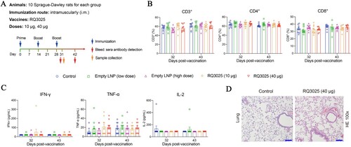
The safety of RQ3025 was evaluated in Sprague–Dawley rats, a strain widely acknowledged as suitable for toxicological research on drugs that have been or are intended to be used in humans [Citation24]. Fifty rats were equally divided into five groups. Each group, on day 0, received intramuscularly one dose of either saline, 10 or 40 μg of RQ3025, or empty LNPs (0.22 mg or 0.86 mg of total lipid) ((A)). All groups received booster injections on days 14 and 28. Neither fever nor weight loss was observed in all animals (Figure S6). Blood samples were collected at various time points for hematological and biochemical analysis. No significant changes were detected in hematological indices or in the percentages of CD3+, CD4+, and CD8+ T cells between the groups (Figure S7 and (B)). The secretions of Th1 cytokines (IFN-γ, TNF-α, and IL-2) and Th2 cytokines (IL-6) were not changed by injections of RQ3025 or empty LNP ((C) and Figure S8). Rats injected with saline or 40 µg of RQ3025 were euthanized on day 31. Lungs, brains, hearts, livers, spleens, and kidneys were harvested and sectioned for H&E staining for histopathological analysis. Vaccine-associated immunopathologic changes were not observed in any of the sections from these tissues in all animals ((D) and Figure S9), suggesting that the administration of RQ3025 at a high dose (40 µg) was safe in rats.
Figure 7. Safety evaluation of RQ3025 in Sprague-Dawley rats. (A) The immunization scheme for Sprague-Dawley (SD) rats. Rats (n = 10 per group) were immunized three times on days 0, 14, and 28 through intramuscular injections with the following substances respectively: Saline (Control), low dose RQ3025 (10 μg), high dose RQ3025 (40 μg), low dose and high dose of empty LNP. Time points of vaccination, bleeding, and sample collection are indicated by arrows. (B, C) Hematological analysis in all five groups of rats. Percentages of lymphocyte subsets including CD3+, CD4+, and CD8+, and expressions of cytokines IFN-γ, TNF-α, and IL-2 were monitored on days indicated. The black dashed line indicates the detection limit of the assay (IFN-γ, 9.40 pg/mL; TNF-α, 3.40 pg/mL; IL-2, 44.10 pg/mL). Any measurement below the detection limit was assigned a value of half the limit of detection for plotting and statistical purposes. Values are mean ± SEM. (D) Histopathological evaluations (H&E staining) in lungs from three groups of rats on day 31. Scale bar, 200 μm. Statistical analyses were performed using ANOVA and Tukey’s multiple comparison tests.
The immunogenicity of RQ3025 in Sprague–Dawley rats was also assessed. Rats immunized with 10 μg and 40 μg doses of RQ3025 induced high titers of IgGs binding to RBD (wild-type), S (wild-type, B.1.351 and BA.5), and S2 (Figure S10A). High titers of neutralizing capacity against B.1.351 and a range of Omicron subvariants (BA.2.3.20, BA.4/BA.5, BQ.1.1, XBB.1, XBB.1.5 and CH.1.1) in the sera of vaccinated rats were demonstrated by a pseudovirus virus neutralization assay (Figure S10B). The seroconversion rate was 100% for B.1.351 and all the tested Omicron subvariants (Figure S10B). These data demonstrate the safety of RQ3025 and its broad-spectrum cross-neutralization activity in rats, supporting its candidacy for clinical development.
Discussion
Amid the COVID-19 pandemic, mRNA vaccine technology demonstrated a great advantage in quick response, contributing largely to the establishment of population-level immunity against SARS-CoV-2. The World Health Organization declared an end to COVID-19 as a global health emergency on 5 May 2023. However, risks remain as the virus continue to evolve and new variants emerge. The development of vaccines that provide broad-spectrum protection is critical to prevent surges in cases and hospitalizations caused by new variants. Here, we present comprehensive assessments of a novel bivalent mRNA vaccine, RQ3025, evaluating its immunogenicity, efficacy, and safety in animal models, providing compelling evidence for its further exploration in clinical trials.
The mRNA vaccines mRNA-1273 and BNT162b2 developed soon after the onset of the COVID-19 pandemic demonstrated over 90% protection efficacy during the initial stages of the pandemic [Citation25]. However, SARS-CoV-2 variants such as Delta and Omicron with immune escape capacity through mutations in the spike protein emerged quickly and significantly reduced the protective efficacy of the vaccines [Citation4,Citation7,Citation10]. It is difficult to predict mutations that can lead to high infectivity and strong immune escape capacity in emerging variants. The vaccine development strategy based on a single strain struggles to keep pace with the rapid evolution of the virus. Various evolutionary paths of SARS-CoV-2 have been identified, and a universal antigen designed based on a comprehensive analysis of evolutionary mutation sites in the spike protein sequences of SARS-CoV-2 variants could effectively induce broad neutralizing antibodies [Citation26,Citation27]. Currently, bivalent mRNA vaccines are widely used to maintain protection efficacy against rapidly emerging SARS-CoV-2 variants, although results were subtle when used as a booster dose [Citation28]. The newly developed bivalent mRNA vaccine RQ3025 presented in this study comprises two mRNA sequences (RQ3013 and RQ3019) encoding chimeric S proteins incorporating key mutation sites found in Alpha, Beta, and Omicron BA.2/BA.4/BA.5 variants of SARS-CoV-2. Most of these mutations exhibited evolutionary conservation and were harbored by numerous recent variants, including BQ.1.1, XBB.1.5, CH.1.1 and XBB.1.16. Our results in various animal models demonstrated that RQ3025 induced high levels of broad neutralizing antibodies against a range of variants including B.1.351, BA.2, BA.2.3.20, BA.2.38, BA.2.75, BA.4, BA.5 BF.7, BQ.1.1, XBB.1, XBB.1.5, XBB.1.16 and CH.1.1, in contrast to the limited neutralizing capacity shown by the monovalent vaccine RQ3013 and RQ3019. With seroconversion rates of 100% for most of these variants, RQ3025 appears to induce relatively unbiased cross-neutralizing antibody responses, attributed to its broad coverage of mutations on the spike protein. Two doses of RQ3025 effectively suppressed the replication of the Omicron variant BA.5 and ameliorated lung injury caused by viral infection in both mice and hamsters. Robust T-cell immune responses persisted for three and two months after the second immunization with RQ3025 in BALB/c and K18-hACE2 mice, respectively, underscoring the vaccine's durable protective efficacy.
In developing vaccines against SARS-CoV-2, concerns about VAERD arose, although it has not been reported in humans receiving current vaccines [Citation1,Citation2]. It has been reported that VAERD in SARS-CoV-2-infected Syrian hamsters immunized with a vaccine consisting of misfolded spike protein and aluminum-containing adjuvants is associated with a Th2-biased immune response1. In our study, a Th1-biased immune response was evident after stimulation with S1 or S2 peptide pools in splenocytes isolated from RQ3025-vaccinated mice, indicating that VAERD is unlikely to develop in RQ3025-vaccinated BALB/c mice.
Currently, the vaccination rate with inactivated COVID-19 vaccines has surpassed 90% in China [Citation29], and a portion of the global population has also received inactivated COVID-19 vaccines. In this study, we evaluated the immunogenicity of different vaccines as booster doses given to BALB/c mice previously immunized with two doses of an inactivated COVID-19 vaccine. The results revealed that a booster dose with RQ3025 generated broader protection with high-titer neutralizing antibodies against B.1.351 and Omicron subvariants (BA.4/BA.5, XBB.1, and BQ.1.1) and induced strong cell-mediated immunity, as shown by a specific T cell immune response against the S protein, demonstrating significant advantages over the inactivated vaccine and monovalent mRNA vaccine RQ3013.
To date, hundreds of SARS-CoV-2 variants have been documented from human patients and the virus continue to evole rapidly. Recently, accelerated evolution of SARS-CoV-2 has been discovered in animal hosts [Citation30]. It is essential to continuously monitor emerging variants and adjust strategies for vaccine development and administration accordingly as new variants may cause serious consequences for human society in the future. The preclinical evaluation of the bivalent mRNA vaccine RQ3025 in this study provides evidences of its excellent broad protective capacity against a range of current variants and supports further exploration of its protective efficacy against current and future variant strains in human.
Disclosure statement
Jinzhong Lin is a co-founder of RNACure and serves on the advisory board of RNACure. The other authors have no conflicts of interest to declare.
Author contributions
J. LIN and J. LU contributed to the conception of the study. J. LIN, J. LU and S.T. designed the experiments. S.T., H.G., W.H., Z. YU, G.L., Z.W., Z. YAO, F.Y., Y.Y., and H.W. participated in multiple experiments. X.G. and J.Z. participated in the production and quality control of mRNA-LNPs. K.L., X.H., M.L., and C.S. conducted live virus challenge experiments involving K18-hACE2 mice and hamsters. S.T., H.G., W.H., J.LU, and J.LIN analyzed the data. W.L., H.Z., and H.Y. helped perform the analysis with constructive discussions. J. LIN, J. LU, S.T., H.G. and W.H. wrote the manuscript. All authors provided the final approval of the manuscript.