Figures & data
Table 1 Effect of acute Zn3P2 poisoning on liver function parameters and hepatic zinc concentration.
Fig. 1 Transmission electron micrograph of the chick’s hepatocyte in control group showing mitochondria (M), rough endoplasmic reticulum (r), glycogen (G), nucleus (N), microvilli (v) and endothelial cell (E). (x2000).
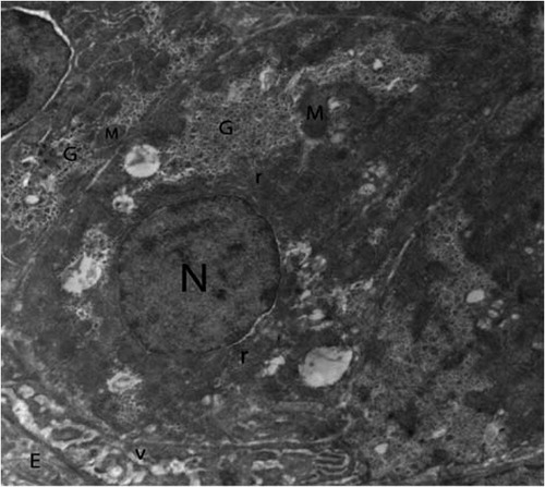
Fig. 2 Higher magnification of control hepatocytes showing several shapes of mitochondria (M), cristae (arrows) rough endoplasmic reticulum (r) and glycogen (G). (×5000).
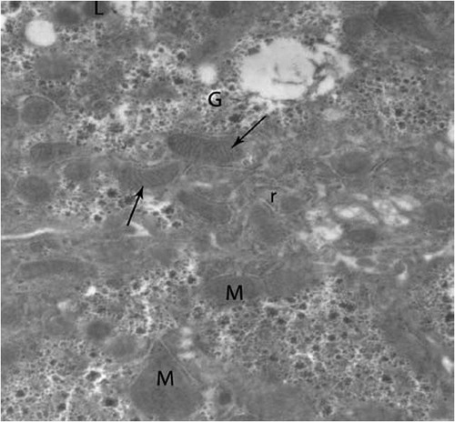
Fig. 3 Electron micrograph of chick’s liver in control group showing hepatocytes (H), mitochondria (M), glycogen (G), kupffer cell (K), erythrocyte (T), endothelial cell (E), lysosomes (L) and lysosomes with electron dense content (arrows) (×2500).
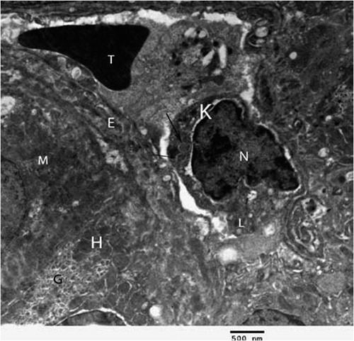
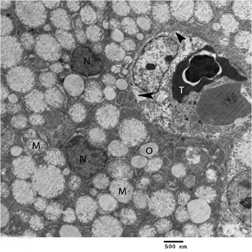
Fig. 4 Transmission electron micrograph of the hepatocyte of chicks intoxicated by zinc phosphide showing swollen mitochondria (M), vacuoles (O), nucleus (N), eryrhrocytes (arrow), intercalated cell (Int) and kupffer cell (K) (×1000).
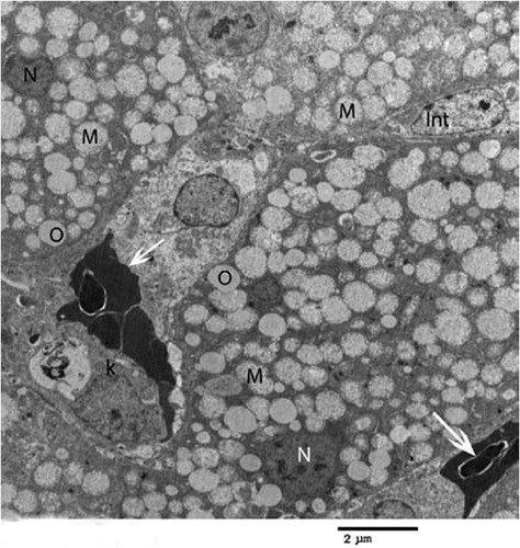
Fig. 5 Higher magnification of intoxicated hepatocyte showing swollen mitochondria (M), cristolysis (arrow), flocculant material (asterisk), dilated rough endoplasmic reticulum (r), glycogen (G), vacuoles (O) and nucleus (N) (×3000).
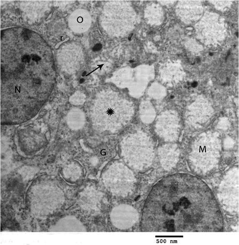
Fig. 6 Another electron micrograph of the hepatocyte of chicks treated with zinc phosphide showing swollen mitochondria (M), vacuoles (O), erythrocyte (T) and shrunken nucleus (N) and absence of microvilli in sinusoids (arrow head) (×2000).