619
Views
1
CrossRef citations to date
0
Altmetric
Research articles

Food bodies of Cecropia pachystachya (Cecropiaceae) leaves: structural and functional features suggesting complementary role to Müllerian bodies

&
Pages 323-334 | Received 04 Sep 2015, Accepted 19 Jan 2016, Published online: 22 May 2016

Figures & data

Figure 1. Food bodies of the leaf of C. pachystachya. A, General aspect of the young leaf before stipule rupture, arrows indicate FBs; B, fully developed FB, note a fragile stalk; C–D, FBs observed under SEM, note the dense indumentum and, in D, the stalk rupture; E–F, detail of the surface of a FB showing thin cuticle and a pore (arrow). Abbreviations: cc, central cells; ep, epidermis; st, stalk.
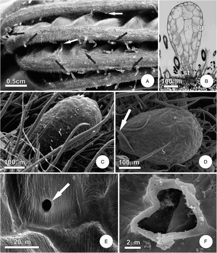
Figure 1. Food bodies of the leaf of C. pachystachya. A, General aspect of the young leaf before stipule rupture, arrows indicate FBs; B, fully developed FB, note a fragile stalk; C–D, FBs observed under SEM, note the dense indumentum and, in D, the stalk rupture; E–F, detail of the surface of a FB showing thin cuticle and a pore (arrow). Abbreviations: cc, central cells; ep, epidermis; st, stalk.

Figure 2. Diagram showing major developmental stages of FBs on the leaf of C. pachystachya. A, Initial stage of FB formation, note the elevation of epidermis due to proliferation of the underlying tissue; B–C, FB at the expansion stage, note the effects of cell expansion and, in C, the persistence of meristematic activity in the stalk cells.
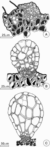
Figure 2. Diagram showing major developmental stages of FBs on the leaf of C. pachystachya. A, Initial stage of FB formation, note the elevation of epidermis due to proliferation of the underlying tissue; B–C, FB at the expansion stage, note the effects of cell expansion and, in C, the persistence of meristematic activity in the stalk cells.

Figure 3. Ultrastructural aspects of FBs of the leaf of C. pachystachya. A–B, Epidermal cell with lamellar outer periclinal wall and organelles in peripheral cytoplasm; C–F, parenchyma of the core of the FBs. In C–D note the cell with large vacuole and lobated nucleus. Detail of peripheral cytoplasm with mitochondria is shown in E–F, note in F a large oil droplet. Abbreviations: di, dictyosome; er, endoplasmic reticulum; mi, mitochondria; nu, nucleus; od, oil droplet; va, vacuole.
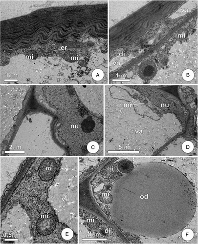
Figure 3. Ultrastructural aspects of FBs of the leaf of C. pachystachya. A–B, Epidermal cell with lamellar outer periclinal wall and organelles in peripheral cytoplasm; C–F, parenchyma of the core of the FBs. In C–D note the cell with large vacuole and lobated nucleus. Detail of peripheral cytoplasm with mitochondria is shown in E–F, note in F a large oil droplet. Abbreviations: di, dictyosome; er, endoplasmic reticulum; mi, mitochondria; nu, nucleus; od, oil droplet; va, vacuole.

Reprints and Corporate Permissions

Please note: Selecting permissions does not provide access to the full text of the article, please see our help page How do I view content?

To request a reprint or corporate permissions for this article, please click on the relevant link below:

Academic Permissions

Please note: Selecting permissions does not provide access to the full text of the article, please see our help page How do I view content?

Obtain permissions instantly via Rightslink by clicking on the button below:

If you are unable to obtain permissions via Rightslink, please complete and submit this Permissions form. For more information, please visit our Permissions help page.