

Figures & data
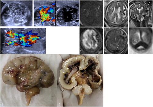
Figure 1. (A, B) Disorganised foetal intracranial structures with abnormal blood flow. Dilated superior vena cava, bilateral cephalic brachial veins and left innominate vein. (C–E, triangle) Multiple patches of high-intensity signals on T1-weighted imaging and low-intensity signals on T2-weighted imaging were distributed around the bilateral lateral ventricles just beneath the ependyma and mainly involved the left germinal matrix. (D, E, arrows) A tortuous and dilated vessel was exposed on the convex surface of the left cerebral hemisphere, and it converged into the superior sagittal sinus along with the surrounding dilated vessels. (F, triangle) The left cerebral hemisphere showed a diffuse high-intensity signal. (G, H, arrows) The bilateral transverse sinuses and sigmoid sinuses were also dilated. (I, arrows) A PAVF in a large vein on the surface of the left hemisphere. (J, arrows) Multiple haemorrhagic foci were found in the germinal matrix. In addition to diffuse encephalomalacia, atrophy of the left cerebral hemisphere and cortical dysplasia were present.
Data availability statement
Data sharing is not applicable to this article as no new data were created or analysed in this study.