Figures & data
Figure 1. Case 1: sagittal T1-W (A) and T2-W (B) images of thoraco-lumbar spinal cord showing a roundish, well-demarcated lesion at the level of the 13th thoracic vertebra (red arrows). The lesion was iso- to slightly hypointense on T1-W images (A) and hyperintense on T2-W. Case 2: T1-W (C) and T2-W transverse (D) views of the apparently intradural-intramedullary cyst located at the level of L1 (yellow arrows). The content of the cyst appeared hyperintense in T2-W and hypointense on T1-W images.
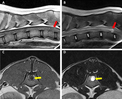
Figure 2. Intraoperative view of the spinal cord in case 1. After durotomy, a grayish white, fluid-filled mass was visualized (arrow).
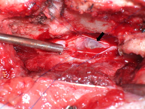
Figure 3. Case 1: (A) histology depicts a cystic and multiloculated, moderately cellular epithelial cell proliferation embedded in moderate amount of stroma, hematoxylin, and eosin stain (2.5×). (B) Higher magnification of (A) columnar epithelium lining an empty cavity and presenting with cilia (arrow head) consistent with respiratory epithelium, hematoxylin, and eosin stain (20×). Case 2: (C) Thin-layered epithelium within the neuroenteric cyst featuring multiple goblet cells containing acidic mucin (arrow head), Alcian blue stain (20×). (D) Immunohistochemistry for pancytokeratin showing a strongly immunoreactive pseudostratified to stratified cuboidal epithelium, DAB as chromogen (20×).
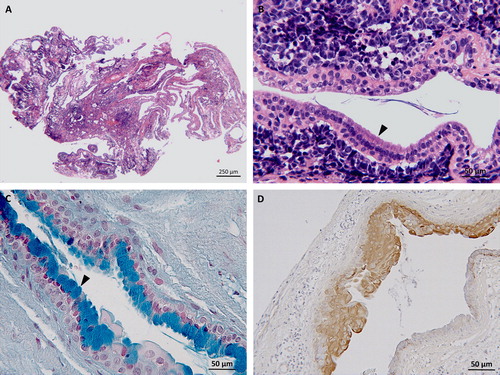
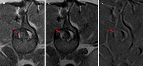
Figure 4. Case 1: Transverse T1-W pre (A) and post-contrast (B) images and subtraction (C) at the level of the surgical site. Note the asymmetrical meningeal thickening and enhancement (red arrow).