Figures & data
Figure 1. Experimental apparatus: An internally-cooled RF electrode (white arrow) has been inserted into an NaCl gel filled well within an Agar phantom, placed in a saline bath at a fixed distance from the grounding pad (G). Thermocouple probes (white curved arrow) have been inserted to measure temperature. An acrylic guide (open arrow) ensures proper positioning of the thermocouple. The RF generator and a temperature measurement device can be seen in the background.
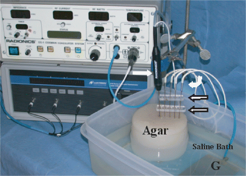
Figure 2. Thermal conductivity as a function of hydrogenated fat concentration. A negative exponential correlation between hydrogenated fat and thermal conductivity (r2 = 0.98) is demonstrated.
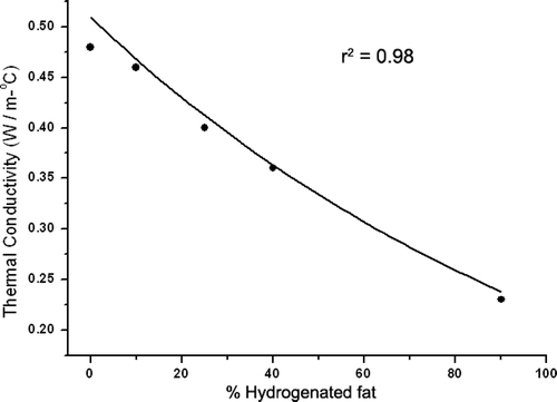
Figure 3. Temperature distribution in agar phantoms subject to RF heating. Temperatures recorded at 1 cm, 2 cm, 3 cm, 4 cm from the electrode show significantly increased temperature at 1 cm (the interface between the 0% fat inner compartment and the background tissue), but slightly lower temperature increase at 2 cm for 90% fat background.
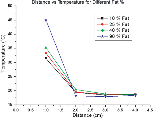
Figure 4. Computer generated temperature profiles of RF heating for varied thermal conductivities. Over a wide range of background tissue thermal conductivities, increased temperatures are observed throughout the 1 cm inner compartment possessing a thermal conductivity of normal tissue (0.5 W m−1°C). For the background tissues, lower temperatures are seen in the presence of lower thermal conductivity. These thermal profiles are similar to those experimentally derived and presented in .
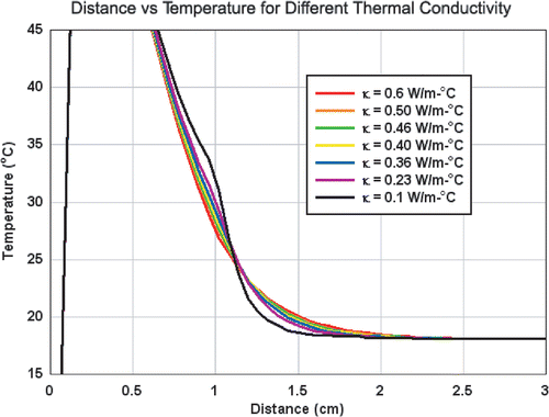
Figure 5. Comparison of experimental and computer generated temperature profiles. Scatter plots of the measured temperatures for the four phantoms (squares) are compared to computer generated simulation data (diamonds). Overall, a tight linear correlation between computer simulations and experimentally generated data was observed (r2 = 0.93).
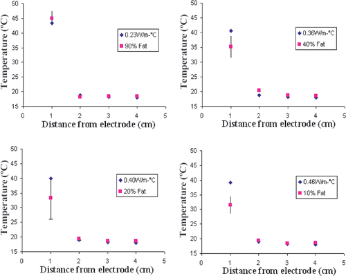
Figure 6. Computer-modelled effect of perfusion on temperatures at the ‘tumour’ margin for tissues with different thermal conductivities with different RF application times. (a) Decreased thermal conductivity within background tissue (i.e. increased lipid content—0.23 W m−1°C for fat and 0.46 W m−1°C for normal soft tissue) can significantly increase the temperature at the margin of tumour in the absence of perfusion. This ‘oven’ effect can be magnified by increased RF ablation time. (b) The presence of perfusion reduces the ‘oven’ effect (i.e. the temperature difference, ΔT°C, between fat and soft tissue background) and the temperature rise at the margin of the tumour, but the range of variability of temperature at the margin of the tumour falls mostly within 50–65°C (depicted by the gray zone), the range of threshold temperatures for inducing necrosis in various tissues Citation[42]. (c) For all durations studied, there is a linear decrease in the ΔT°C at the tumour margin relative to increasing tissue perfusion (r2 = 0.99). Additionally, the negative slopes of ΔT°C for increasing perfusion is greater for longer RF durations (6–24 min).
![Figure 6. Computer-modelled effect of perfusion on temperatures at the ‘tumour’ margin for tissues with different thermal conductivities with different RF application times. (a) Decreased thermal conductivity within background tissue (i.e. increased lipid content—0.23 W m−1°C for fat and 0.46 W m−1°C for normal soft tissue) can significantly increase the temperature at the margin of tumour in the absence of perfusion. This ‘oven’ effect can be magnified by increased RF ablation time. (b) The presence of perfusion reduces the ‘oven’ effect (i.e. the temperature difference, ΔT°C, between fat and soft tissue background) and the temperature rise at the margin of the tumour, but the range of variability of temperature at the margin of the tumour falls mostly within 50–65°C (depicted by the gray zone), the range of threshold temperatures for inducing necrosis in various tissues Citation[42]. (c) For all durations studied, there is a linear decrease in the ΔT°C at the tumour margin relative to increasing tissue perfusion (r2 = 0.99). Additionally, the negative slopes of ΔT°C for increasing perfusion is greater for longer RF durations (6–24 min).](/cms/asset/1da05339-467c-4148-a3fd-b0192fd525be/ihyt_a_160895_f0006_b.gif)
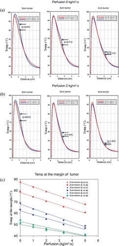
Figure 7. Computer-modelled effect of perfusion on temperatures at the ‘tumour’ margin for tissues with different thermal conductivities with different tumour sizes. Decreased thermal conductivity within background tissue (i.e. increased lipid content—0.23 W m−1°C for fat and 0.46 W m−1°C for normal soft tissue) can significantly increase the temperature difference (ΔT°C) at the margin of tumour in the absence of perfusion (a) and with a perfusion of 2 kg m−3 s−1 (b). This ‘oven’ effect is diminished as tumour diameter increases. (c) Furthermore, there is a decreasing linear relationship between increasing perfusion and temperatures at the margin at the interface of tumour and surrounding tissue for all tumour sizes (2–4 cm in diameter) (r2 = 0.94–0.99).