Figures & data
Figure 1. Diagram of the layers in the multilayer conformal applicator, including thermal monitoring catheters, a waterbolus, a printed circuit board microwave array, a dielectric spacer, brachytherapy catheters, and an inflatable air bladder. The stretch fabric is a separate elastic garment that may be worn over the applicator.
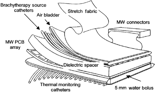
Figure 2. Sequential diagrams of an ‘L-shaped’ multilayer conformal applicator showing the different layers. (a) Rear view of the applicator showing elastic straps and skin-contacting thermometry catheters; (b) front view of the water bolus; (c) PCB array; (d) dielectric spacer; (e) brachytherapy catheters; (f) inflatable air bladder.
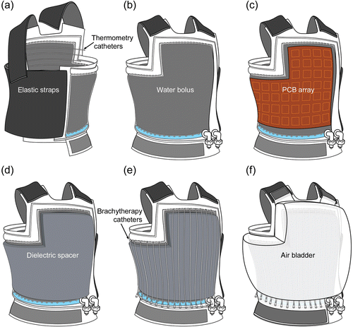
Figure 3. Design diagrams for waterboluses showing water ports, channels, and numbered thermocouple locations used in thermometry evaluation of fluid flow dynamics. (a) Rectangular bolus with a 19 × 34 cm treatment area; (b) ‘L’ shaped bolus with a 32 × 42 cm treatment area.
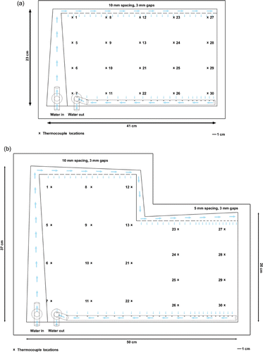
Figure 4. Human torso CT phantom fitted with a conformal ‘L’ applicator in preparation for CT imaging and evaluation of applicator conformity. (a) Waterbolus layer fitted on phantom with elastic straps; (b) complete applicator fitted on phantom with inflated air bladder and an elastic garment.
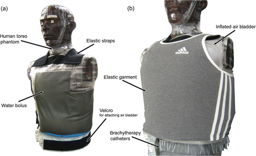
Figure 5. Axial CT cross-sections from the upper torso of a CT phantom fitted with the multilayer conformal applicator. (a) Axial cross-section of the upper torso prior to air bladder inflation; (b) close-up of the bolus over the breast and sternum prior to air bladder inflation; (c) axial cross section of the upper torso after air bladder inflation; (d) close-up of the bolus over the breast and sternum after air bladder inflation.
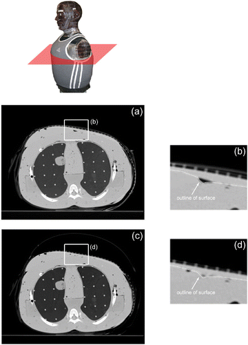
Figure 6. Axial CT cross-sections from the lower torso of a CT phantom fitted with the multilayer conformal applicator. (a) Axial cross-section of lower torso prior to air bladder inflation; (b) close-up of bolus over breast and sternum prior to air bladder inflation; (c) close-up of small air pockets under the arm prior to air bladder inflation; (d) axial cross-section of lower torso after air bladder inflation; (e) close-up of bolus over breast and sternum after air bladder inflation; (f) close-up of reduced air pockets under the arm after air bladder inflation; (g) axial cross-section of lower torso after simulated patient movement.
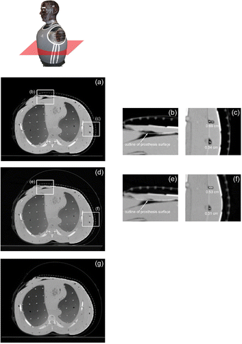
Figure 7. Time temperature plots of the surface temperatures measured at the numbered thermocouple locations indicated in . (a) Rectangular bolus with a 19 × 34 cm treatment area, water flow rate at 1.54 min; (b) "L" shaped bolus with a 32 × 42 cm treatment area, water flow rate at 2.4 L/min.
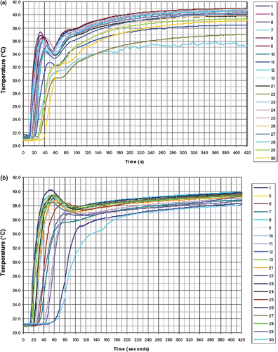
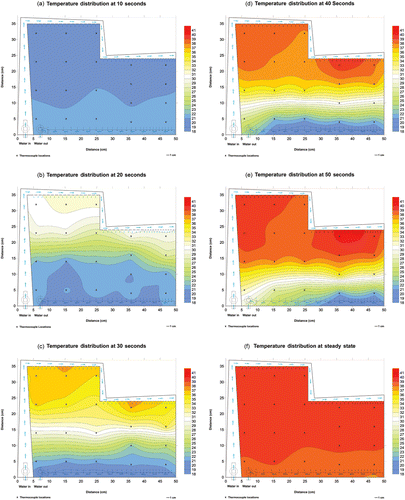
Figure 8. Color wash images displaying changing temperature distributions across the ‘L’ shaped bolus from 10–50 s and at steady state. (a) 10 s; (b) 20 s; (c) 30 s; (d) 40 s; (e) 50 s; (f) steady state.