1,266
Views
47
CrossRef citations to date
0
Altmetric
Original

Hemorrhage control using high intensity focused ultrasound

, PhD &
Pages 203-211 | Received 16 Sep 2006, Accepted 01 Dec 2006, Published online: 09 Jul 2009

Figures & data

Figure 1. Protocol for hemorrhage control using image-guided HIFU.

Figure 1. Protocol for hemorrhage control using image-guided HIFU.

Figure 2. Gross (a) and light microscopy (b) observations of a HIFU-sealed puncture site in the femoral artery. The puncture was sealed by coagulated blood (white arrow) and a fibrous cap (black arrow). The histological slide was stained with Masson's trichrome stain.
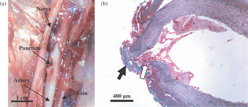
Figure 2. Gross (a) and light microscopy (b) observations of a HIFU-sealed puncture site in the femoral artery. The puncture was sealed by coagulated blood (white arrow) and a fibrous cap (black arrow). The histological slide was stained with Masson's trichrome stain.

Figure 3. (a) HIFU annular array, integrated with an ultrasound imaging probe (arrow). (b) Before HIFU, patent hepatic vein (outlined by dashed lines) is visualized using Color Doppler (arrow). (c) During HIFU, hyperecho (arrow) formed at the position of the vessel. (d) After HIFU, the occluded vein (outlined by dashed lines) shows no flow (arrow). The site of treatment is seen as a hyperechoic region surrounding a vein that appears to be collapsed (arrowhead).

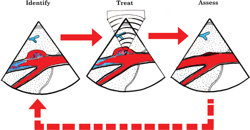
Figure 3. (a) HIFU annular array, integrated with an ultrasound imaging probe (arrow). (b) Before HIFU, patent hepatic vein (outlined by dashed lines) is visualized using Color Doppler (arrow). (c) During HIFU, hyperecho (arrow) formed at the position of the vessel. (d) After HIFU, the occluded vein (outlined by dashed lines) shows no flow (arrow). The site of treatment is seen as a hyperechoic region surrounding a vein that appears to be collapsed (arrowhead).

Figure 4. Pulsed Doppler spectrum obtained from the femoral artery before and after the injury was produced. (a) Normal arterial blood flow before the injury. (b) Bleeding artery shows a spectrum with increased baseline and elevated peak amplitude.

Figure 4. Pulsed Doppler spectrum obtained from the femoral artery before and after the injury was produced. (a) Normal arterial blood flow before the injury. (b) Bleeding artery shows a spectrum with increased baseline and elevated peak amplitude.

Reprints and Corporate Permissions

Please note: Selecting permissions does not provide access to the full text of the article, please see our help page How do I view content?

To request a reprint or corporate permissions for this article, please click on the relevant link below:

Academic Permissions

Please note: Selecting permissions does not provide access to the full text of the article, please see our help page How do I view content?

Obtain permissions instantly via Rightslink by clicking on the button below:

If you are unable to obtain permissions via Rightslink, please complete and submit this Permissions form. For more information, please visit our Permissions help page.