1,292
Views
45
CrossRef citations to date
0
Altmetric
Original

Interstitial devices for minimally invasive thermal ablation by high-intensity ultrasound

, , &
Pages 153-163 | Received 14 Nov 2006, Accepted 14 Dec 2006, Published online: 09 Jul 2009

Figures & data

Figure 1. Transducers for interstitial applications: (a) Tip of a dual mode intracardiac ablation tool. The applicator consists in a 112-element imaging array surround by a coagulating ring (Courtesy of KL Gentry). (b) Multi-element cylindrical transducers for coagulation in prostate under MR guidance (Courtesy of CJ Diederich and WH Nau). (c) 64-element cylindrical array for treating oesophageal tumours. Plane waves are reconstructed and rotated electronically. (d) Intracardiac catheter producing focused coagulation around the pulmonary vein for treating atrial fibrilation (Courtesy of DA Smith, ProRhythm, Inc.).
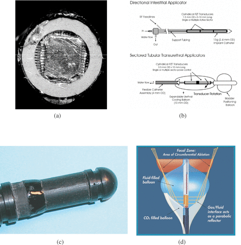
Figure 1. Transducers for interstitial applications: (a) Tip of a dual mode intracardiac ablation tool. The applicator consists in a 112-element imaging array surround by a coagulating ring (Courtesy of KL Gentry). (b) Multi-element cylindrical transducers for coagulation in prostate under MR guidance (Courtesy of CJ Diederich and WH Nau). (c) 64-element cylindrical array for treating oesophageal tumours. Plane waves are reconstructed and rotated electronically. (d) Intracardiac catheter producing focused coagulation around the pulmonary vein for treating atrial fibrilation (Courtesy of DA Smith, ProRhythm, Inc.).

Figure 2. (a) Temperature map acquired in vivo during an ultrasound exposure using interstitial applicator. The applicator is positioned in the oesophagus. The absolute temperature scale 42–47°C for the regions shown in blue, 48–52°C for regions in green, and above 53°C for regions in red. (b) Fluoroscopic image for positioning an endoscopic ultrasound applicator in the bile duct and treating cholangiocarcinoma. 1–Ultrasound therapeutic transducer, 2–Applicator, 3–Guide wire, 4–Endoscope.
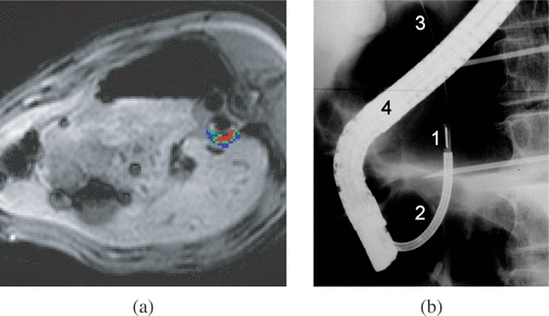
Figure 2. (a) Temperature map acquired in vivo during an ultrasound exposure using interstitial applicator. The applicator is positioned in the oesophagus. The absolute temperature scale 42–47°C for the regions shown in blue, 48–52°C for regions in green, and above 53°C for regions in red. (b) Fluoroscopic image for positioning an endoscopic ultrasound applicator in the bile duct and treating cholangiocarcinoma. 1–Ultrasound therapeutic transducer, 2–Applicator, 3–Guide wire, 4–Endoscope.

Reprints and Corporate Permissions

Please note: Selecting permissions does not provide access to the full text of the article, please see our help page How do I view content?

To request a reprint or corporate permissions for this article, please click on the relevant link below:

Academic Permissions

Please note: Selecting permissions does not provide access to the full text of the article, please see our help page How do I view content?

Obtain permissions instantly via Rightslink by clicking on the button below:

If you are unable to obtain permissions via Rightslink, please complete and submit this Permissions form. For more information, please visit our Permissions help page.