19,611
Views
516
CrossRef citations to date
0
Altmetric
Original

High intensity focused ultrasound: Physical principles and devices

&
Pages 89-104 | Received 22 Dec 2006, Accepted 24 Dec 2006, Published online: 09 Jul 2009

Figures & data

Figure 1. Diagrammatic illustration of the principle of HIFU. High-intensity ultrasound waves are generated by a transducer outside the body and focussed onto a small region deep within tissue. HIFU-induced heating causes cell death by thermal necrosis within the focal volume, but leaves tissue elsewhere in the propagation path unaffected.
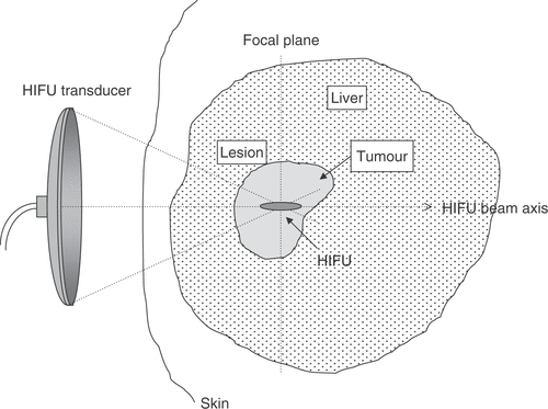
Figure 1. Diagrammatic illustration of the principle of HIFU. High-intensity ultrasound waves are generated by a transducer outside the body and focussed onto a small region deep within tissue. HIFU-induced heating causes cell death by thermal necrosis within the focal volume, but leaves tissue elsewhere in the propagation path unaffected.

Table I.  Characteristics of transducers in use for clinical applications of HIFU.

Figure 2. Superposition onto the HIFU treatment geometry of a typical axial HIFU pressure profile (a) and of a diagrammatic representation of the frequency-content, f, of the HIFU wave (b–d) as it propagates through tissue. (a) Representative axial pressure distribution (characterised using a hydrophone in water) for a typical HIFU transducer. The large peak defines the focal region of the HIFU transducer, within which thermal damage is expected to occur. Prefocal peaks can also be seen to exist which, should they overlap with a tissue region that is either highly nonlinear or has a high absorption coefficient, are likely to cause significant prefocal heating. The HIFU transducer is generally excited sinusoidally with a single frequency, f0, resulting in a monochromatic wave (b). As this wave travels through a nonlinear medium, superharmonic leakage occurs (c) and energy at these higher harmonics is readily absorbed and converted into heat. Finally, if inertial cavitation occurs (generally, but not necessarily, in the focal region), the collapsing microbubbles convert part of the incident energy into broadband noise emissions (d), which are very rapidly and very locally absorbed and converted into heat.
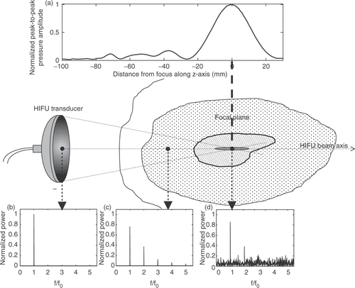
Figure 2. Superposition onto the HIFU treatment geometry of a typical axial HIFU pressure profile (a) and of a diagrammatic representation of the frequency-content, f, of the HIFU wave (b–d) as it propagates through tissue. (a) Representative axial pressure distribution (characterised using a hydrophone in water) for a typical HIFU transducer. The large peak defines the focal region of the HIFU transducer, within which thermal damage is expected to occur. Prefocal peaks can also be seen to exist which, should they overlap with a tissue region that is either highly nonlinear or has a high absorption coefficient, are likely to cause significant prefocal heating. The HIFU transducer is generally excited sinusoidally with a single frequency, f0, resulting in a monochromatic wave (b). As this wave travels through a nonlinear medium, superharmonic leakage occurs (c) and energy at these higher harmonics is readily absorbed and converted into heat. Finally, if inertial cavitation occurs (generally, but not necessarily, in the focal region), the collapsing microbubbles convert part of the incident energy into broadband noise emissions (d), which are very rapidly and very locally absorbed and converted into heat.

Figure 3. Diagrammatic illustration of HIFU treatment delivery for the ablation of large tissue volumes. Multiple lesions are created side-by-side to span the required treatment volume, starting on the side distal to the transducer. The lesions can be overlapping or separate, depending on the necessity to achieve confluent regions of cell killing.
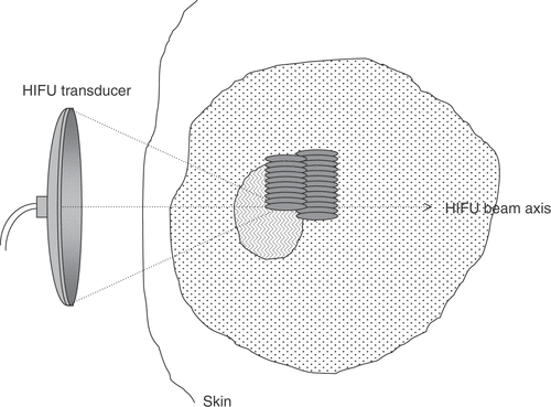
Figure 3. Diagrammatic illustration of HIFU treatment delivery for the ablation of large tissue volumes. Multiple lesions are created side-by-side to span the required treatment volume, starting on the side distal to the transducer. The lesions can be overlapping or separate, depending on the necessity to achieve confluent regions of cell killing.

Reprints and Corporate Permissions

Please note: Selecting permissions does not provide access to the full text of the article, please see our help page How do I view content?

To request a reprint or corporate permissions for this article, please click on the relevant link below:

Academic Permissions

Please note: Selecting permissions does not provide access to the full text of the article, please see our help page How do I view content?

Obtain permissions instantly via Rightslink by clicking on the button below:

If you are unable to obtain permissions via Rightslink, please complete and submit this Permissions form. For more information, please visit our Permissions help page.