Figures & data
Figure 1. Water coupled system. (a) Cylindrical phantoms (5) were held in a tank of degassed water at the focal distance of the single focus transducer (4). Temperature of degassed water was monitored (1) and held constant. Temperature elevations were monitored by thermal couples in controls (no exposure) (2) and HIFU exposed phantoms (3). (b) Mice were anesthetized and held in the tank in the same manner as phantoms. Similarly, temperature elevations by thermal couples were monitored in controls (no exposure) (2) and HIFU exposed tumors (3).
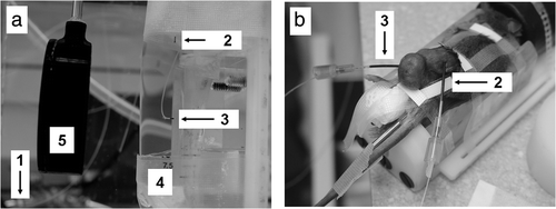
Figure 2. Membrane coupled system. Both single and split focus transducers were housed in a sealed chamber of degassed water. Planar phantoms (3) were placed on the membrane (2) and held at the focal distance of both transducers (1).
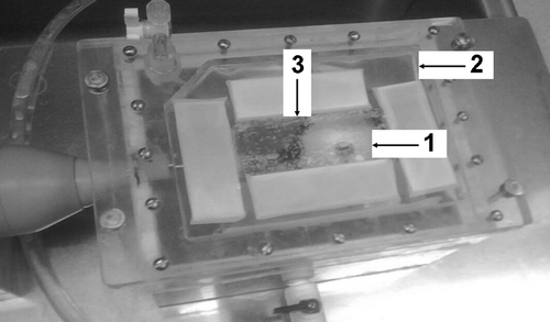
Table I. Experimental exposures.
Figure 3. (a) Temperature elevation in cylindrical phantoms at 80W compared to tumor tissue at 20W. A significant difference was not found between the two. At 20W, temperature elevations in the phantoms were not sufficiently high to be determined accurately. Columns, mean (n = 5); bars, standard deviation. (b) Linear relationship between duty cycle (%) and peak temperature elevation in cylindrical phantoms (80W). Points, mean (n = 5); bars, standard deviation.
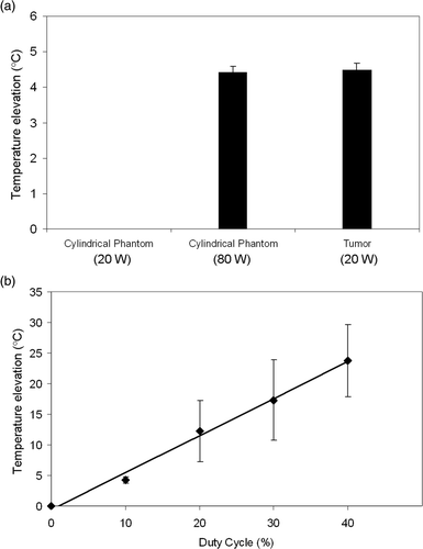
Figure 4. Spatial temperature elevation greater than 3°C and 5°C during HIFU exposure with single focus and split focus transducers. Points, mean (n = 7); bars, standard deviation.
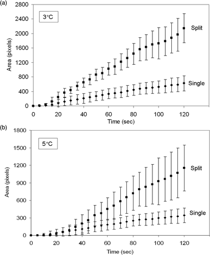
Table II. Single to split focus comparisons.
Figure 5. Area under the curve for spatial temperature elevation greater than 3°C and 5°C during HIFU exposure with single focus and split focus transducers. Columns, mean (n = 7); bars, standard deviation.
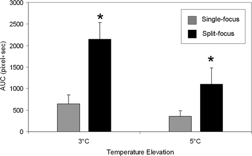
Figure 6. Representative IR heating images for treatment of planar phantoms with single focus and split focus transducers, for a 120s exposure with each device. Images at early time points for the split focus device allow individual foci to be observed, which eventually meld together into a single larger focal spot (compared to the single focus device) by the end of the exposure. (Scale bar = 1 cm).
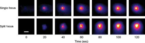
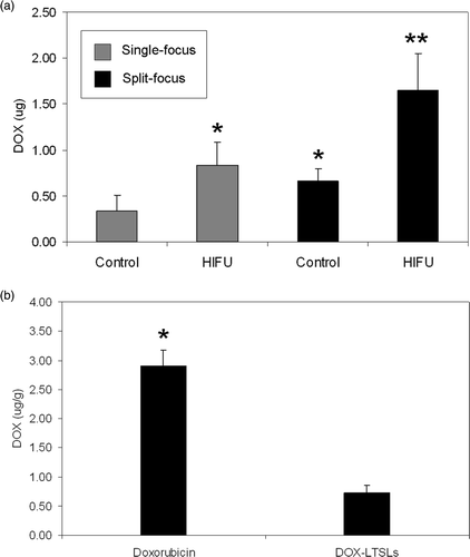
Figure 7. (a) Total doxorubicin extracted from muscle in HIFU exposed extremity and control (unexposed) extremity for single focus and split focus transducers. (b) Concentration of doxorubicin found in muscle following treatment with free doxorubicin and LTSL-doxorubicin. No HIFU exposure. Columns, mean (n = 5); bars, standard deviation.