450
Views
17
CrossRef citations to date
0
Altmetric
Original

RF ablation with adjuvant therapy: Comparison of external beam radiation and liposomal doxorubicin on ablation efficacy in an animal tumor model

, , , , &
Pages 560-567 | Received 08 Nov 2007, Accepted 19 Mar 2008, Published online: 09 Jul 2009

Figures & data

Figure 1. Rat tumor model. This figure demonstrates the experimental set up of RF ablation within the R3230 breast tumor model where (A) illustrates the grounding pad, (B) the RF generator, and (C) denotes the tumor with RF needle inserted. Inset: This illustrates the RF electrode A and thermocouple B inserted into an acrylic marker for insertion at specific radii to enable thermal mapping.
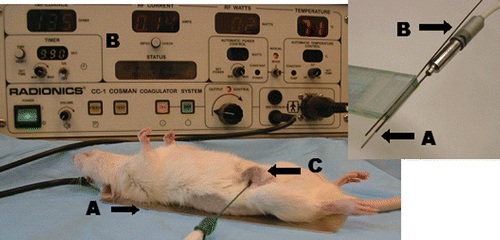
Figure 1. Rat tumor model. This figure demonstrates the experimental set up of RF ablation within the R3230 breast tumor model where (A) illustrates the grounding pad, (B) the RF generator, and (C) denotes the tumor with RF needle inserted. Inset: This illustrates the RF electrode A and thermocouple B inserted into an acrylic marker for insertion at specific radii to enable thermal mapping.

Figure 2. Cross section of stained ablated tumor. This figure demonstrates a cross section of RF ablated tumor stained with TTC. Sharp demarcation is noted at the margin (arrow) between the white, ablated tissue and the unablated surrounding tumor tissue stained pink. Central dot in the white coagulation denotes the original position of the RF electrode.
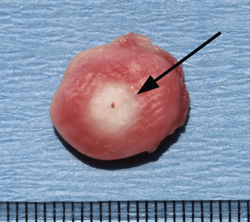
Figure 2. Cross section of stained ablated tumor. This figure demonstrates a cross section of RF ablated tumor stained with TTC. Sharp demarcation is noted at the margin (arrow) between the white, ablated tissue and the unablated surrounding tumor tissue stained pink. Central dot in the white coagulation denotes the original position of the RF electrode.

Table I.  The effects of application duration upon combined adjuvant therapies. This table demonstrates that an increase in RF dose produces increases in coagulation, AUC, and CEM43. In comparison to RF alone and RF combined with Doxil, high dose XRT produces increases in coagulation while decreasing AUC and CEM43 (percentage decreases in AUC are based upon the baseline of RF alone for a given duration). Within each experimental group, duration of application had no effect on temperature threshold for ablation.

Figure 3. Thermal mapping of temperature distributions within a subcutaneous tumor. Mean temperature profiles for multiple measurements at five distances within the tumor (1.5–7 mm from the electrode) are presented with T-bars. The temperatures increase in a sigmoidal Boltzmann distribution (R2 = 0.94–0.99).
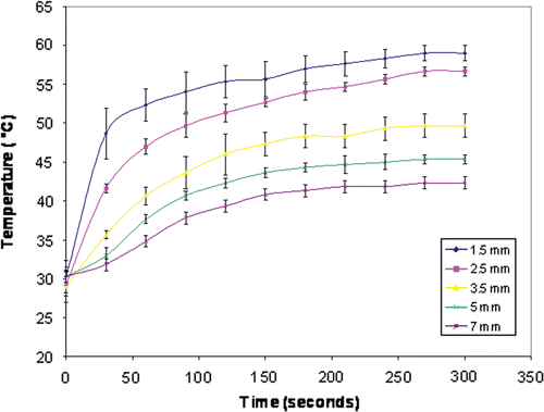
Figure 3. Thermal mapping of temperature distributions within a subcutaneous tumor. Mean temperature profiles for multiple measurements at five distances within the tumor (1.5–7 mm from the electrode) are presented with T-bars. The temperatures increase in a sigmoidal Boltzmann distribution (R2 = 0.94–0.99).

Reprints and Corporate Permissions

Please note: Selecting permissions does not provide access to the full text of the article, please see our help page How do I view content?

To request a reprint or corporate permissions for this article, please click on the relevant link below:

Academic Permissions

Please note: Selecting permissions does not provide access to the full text of the article, please see our help page How do I view content?

Obtain permissions instantly via Rightslink by clicking on the button below:

If you are unable to obtain permissions via Rightslink, please complete and submit this Permissions form. For more information, please visit our Permissions help page.