755
Views
15
CrossRef citations to date
0
Altmetric
Original

Hyperthermia on mesenchymal stem cells (MSCs) can sensitize tumor cells to undergo cell death

, , , &
Pages 638-648 | Received 21 Dec 2007, Accepted 04 Jun 2008, Published online: 09 Jul 2009

Figures & data

Figure 1. Microscopic analysis of untreated and heat-treated MSCs. MSCs were untreated (no shock) or were heat-treated at 43°C for 45 min, followed by incubation at 37°C for 24 h (heat shock). Microscopic observation was performed by phase-contrast microscopy with 10 (left) or 40 (right) fold of magnification. A representative image is displayed from three independent experiments.
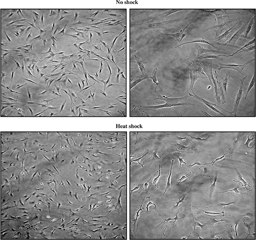
Figure 1. Microscopic analysis of untreated and heat-treated MSCs. MSCs were untreated (no shock) or were heat-treated at 43°C for 45 min, followed by incubation at 37°C for 24 h (heat shock). Microscopic observation was performed by phase-contrast microscopy with 10 (left) or 40 (right) fold of magnification. A representative image is displayed from three independent experiments.

Figure 2. Immunophenotypes of normal and heat-treated MSCs. PE-conjugated anti-CD45, -CD44, -CD90, -CD34, or -CD105 antibodies were reacted with MSCs that were untreated (A, no shock) or incubated for 24 h after heat shock (B, heat shock). The result was obtained by analysis using flow cytometry. A representative image is displayed from three independent experiments.
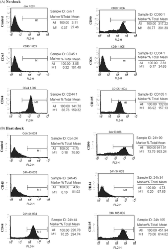
Figure 2. Immunophenotypes of normal and heat-treated MSCs. PE-conjugated anti-CD45, -CD44, -CD90, -CD34, or -CD105 antibodies were reacted with MSCs that were untreated (A, no shock) or incubated for 24 h after heat shock (B, heat shock). The result was obtained by analysis using flow cytometry. A representative image is displayed from three independent experiments.

Figure 3. Proliferation of MCF7 cells cultured in the conditioned medium obtained from non-treated or heat-treated MSCs. MCF7 were incubated in the conditioned medium obtained from MSCs that were non-shocked (no shock) or heat-shocked (heat shock). (A) Cellular morphology was observed by phase-contrast microscopy after incubation for the indicated time (24, 48 or 72 h). Bars; 50 µm. (B) Cell proliferation was measured with Wst-1 reagent by reading the optical density at 450 nm after incubation for the indicated time (24, 48 or 72 h). The experiments were performed three times and demonstrated similar results. Statistical significance: *. P < 0.05.
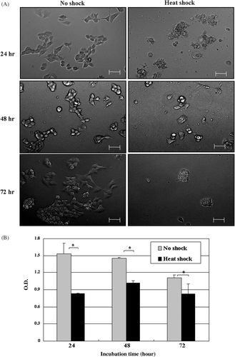
Figure 3. Proliferation of MCF7 cells cultured in the conditioned medium obtained from non-treated or heat-treated MSCs. MCF7 were incubated in the conditioned medium obtained from MSCs that were non-shocked (no shock) or heat-shocked (heat shock). (A) Cellular morphology was observed by phase-contrast microscopy after incubation for the indicated time (24, 48 or 72 h). Bars; 50 µm. (B) Cell proliferation was measured with Wst-1 reagent by reading the optical density at 450 nm after incubation for the indicated time (24, 48 or 72 h). The experiments were performed three times and demonstrated similar results. Statistical significance: *. P < 0.05.

Figure 4. Cell cycle analysis of MCF7 cells which were cultured under the conditioned medium from no-shocked or heat-shocked MSCs. MCF7 cells were incubated in the conditioned medium obtained from MSCs that were no-shocked (No shock) or heat-shocked (Heat shock). After incubation for 24, 48 or 72 h, the cells were stained with propidium iodide solution. The image obtained by analysis using flow cytometry was displayed with the percentage at each phase of cell cycle. Similar data were obtained from two independent experiments.
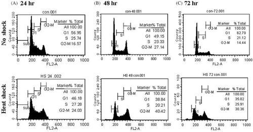
Figure 4. Cell cycle analysis of MCF7 cells which were cultured under the conditioned medium from no-shocked or heat-shocked MSCs. MCF7 cells were incubated in the conditioned medium obtained from MSCs that were no-shocked (No shock) or heat-shocked (Heat shock). After incubation for 24, 48 or 72 h, the cells were stained with propidium iodide solution. The image obtained by analysis using flow cytometry was displayed with the percentage at each phase of cell cycle. Similar data were obtained from two independent experiments.

Figure 5. Analysis of molecular changes of MCF7 cells which were cultured in the conditioned medium from non-shocked or heat-shocked MSCs. MCF7 cells were incubated in the conditioned medium obtained from MSCs that were non-shocked (no shock) or heat-shocked (heat shock). (A) Flow cytometric analysis of surface proteins. MCF7 cells cultured for 48 h were stained either with anti-MHC class I molecules or anti-FAS antibodies, and subsequent PE-conjugated secondary antibodies. The mean fluorescence intensity was obtained by flow cytometry and converged into the graph. One representative result is displayed from two independent experiments. (B) Analysis of mRNA expressions for tumor-associated molecules by RT-PCR. MCF7 cells cultured in each condition for 24, 48 or 72 h were collected to extract RNA which was subsequently reverse-transcribed to cDNA. PCR was performed with each pair of the indicated primers and β-actin as a control. The image was obtained using gel documentation system after electrophoresis and staining the gel with ethidium bromide. Similar data were obtained from two independent experiments.
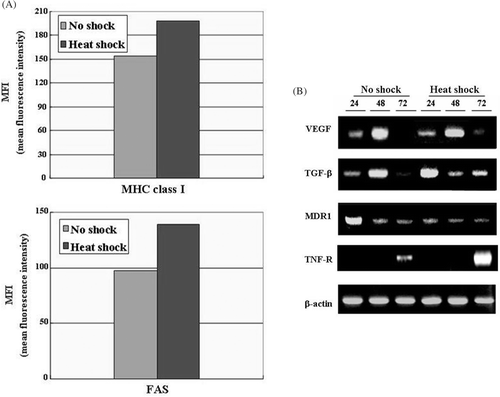
Figure 5. Analysis of molecular changes of MCF7 cells which were cultured in the conditioned medium from non-shocked or heat-shocked MSCs. MCF7 cells were incubated in the conditioned medium obtained from MSCs that were non-shocked (no shock) or heat-shocked (heat shock). (A) Flow cytometric analysis of surface proteins. MCF7 cells cultured for 48 h were stained either with anti-MHC class I molecules or anti-FAS antibodies, and subsequent PE-conjugated secondary antibodies. The mean fluorescence intensity was obtained by flow cytometry and converged into the graph. One representative result is displayed from two independent experiments. (B) Analysis of mRNA expressions for tumor-associated molecules by RT-PCR. MCF7 cells cultured in each condition for 24, 48 or 72 h were collected to extract RNA which was subsequently reverse-transcribed to cDNA. PCR was performed with each pair of the indicated primers and β-actin as a control. The image was obtained using gel documentation system after electrophoresis and staining the gel with ethidium bromide. Similar data were obtained from two independent experiments.

Figure 6. Combined effects of the conditioned medium from non-shocked or heat-shocked MSCs with chemotherapeutic drugs. MCF7 cells were cultured in the conditions containing each indicated drug (5-aza-cytidine, 5-FU, cisplatin, genistein) either with media only or the conditioned medium obtained from MSCs that were non-shocked (no shock) or heat-shocked (heat shock). After 48 h incubation, the cell proliferation was assayed with Wst-1 reagent, and the optical density at 450 nm was displayed. The experiments were performed three times and demonstrated similar results. Statistical significance: **. P < 0.01.
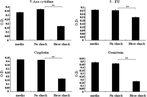
Figure 6. Combined effects of the conditioned medium from non-shocked or heat-shocked MSCs with chemotherapeutic drugs. MCF7 cells were cultured in the conditions containing each indicated drug (5-aza-cytidine, 5-FU, cisplatin, genistein) either with media only or the conditioned medium obtained from MSCs that were non-shocked (no shock) or heat-shocked (heat shock). After 48 h incubation, the cell proliferation was assayed with Wst-1 reagent, and the optical density at 450 nm was displayed. The experiments were performed three times and demonstrated similar results. Statistical significance: **. P < 0.01.
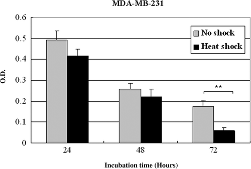
Supplementary Figure 1 Proliferation of MDA-MB-231 cells cultured in the conditioned medium obtained from non-treated or heat-treated MSCs. MDA-MB-231 were incubated in the conditioned medium obtained from MSCs that were non-shocked (no shock) or heat-shocked (heat shock). Cell proliferation was measured with Wst-1 reagent by reading the optical density at 450 nm after incubation for the indicated time (24, 48 or 72 h). The experiments were performed three times and demonstrated similar results. Statistical significance: **. P < 0.01.

Supplementary Figure 1 Proliferation of MDA-MB-231 cells cultured in the conditioned medium obtained from non-treated or heat-treated MSCs. MDA-MB-231 were incubated in the conditioned medium obtained from MSCs that were non-shocked (no shock) or heat-shocked (heat shock). Cell proliferation was measured with Wst-1 reagent by reading the optical density at 450 nm after incubation for the indicated time (24, 48 or 72 h). The experiments were performed three times and demonstrated similar results. Statistical significance: **. P < 0.01.

Supplementary Figure 2 Proliferation of MCF7 and MDA-MB-231 cells cultured in the conditioned medium obtained from non-treated or heat-treated fibroblasts. MCF7 or MDA-MB-231 were incubated in the conditioned medium obtained from primary fibroblast cells that were non-shocked (no shock) or heat-shocked (heat shock). Cell proliferation was measured with Wst-1 reagent by reading the optical density at 450 nm after incubation for the indicated time (24, 48 or 72 h). The experiments were performed three times and demonstrated similar results.
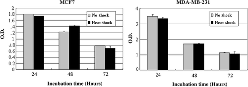
Supplementary Figure 2 Proliferation of MCF7 and MDA-MB-231 cells cultured in the conditioned medium obtained from non-treated or heat-treated fibroblasts. MCF7 or MDA-MB-231 were incubated in the conditioned medium obtained from primary fibroblast cells that were non-shocked (no shock) or heat-shocked (heat shock). Cell proliferation was measured with Wst-1 reagent by reading the optical density at 450 nm after incubation for the indicated time (24, 48 or 72 h). The experiments were performed three times and demonstrated similar results.

Reprints and Corporate Permissions

Please note: Selecting permissions does not provide access to the full text of the article, please see our help page How do I view content?

To request a reprint or corporate permissions for this article, please click on the relevant link below:

Academic Permissions

Please note: Selecting permissions does not provide access to the full text of the article, please see our help page How do I view content?

Obtain permissions instantly via Rightslink by clicking on the button below:

If you are unable to obtain permissions via Rightslink, please complete and submit this Permissions form. For more information, please visit our Permissions help page.