Figures & data
Table I. Survival and apoptosis rates in SAS/neo and SAS/mp53 cells after heating (44°C, 40 min) Citation[15].
Figure 1. Unsupervised hierarchical clustering analysis of 512 proteins in heat treated cells compared with non-treated cells. The graph indicates relative protein expression levels. Lower signal values are indicated by green, and higher signal values are indicated by red.
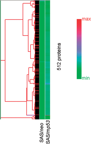
Figure 2. Hierarchical clustering analysis of 44 selected apoptosis-related proteins which showed valid changes in heat treated cells when compared to non-treated cells. The graph indicates protein expression levels with lower signal values shown in green and higher signal values shown in red.
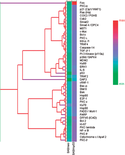
Figure 3. INRs for apoptosis-related proteins which were obtained from protein microarray analysis. The central line is the average value for INRs as measured from the expression of 44 selected apoptosis-related proteins which showed valid changes in response to heat treatment.
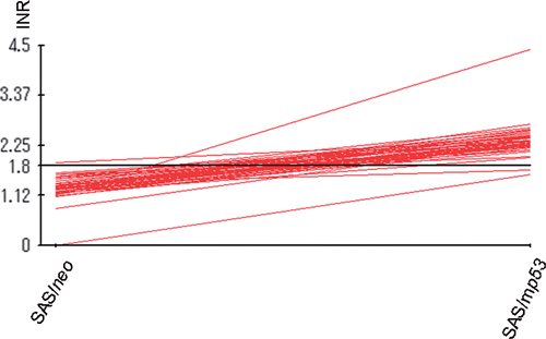
Table II. Apoptosis-related proteins with more than a 1.5-fold increase in expression in SAS/neo cells.
Table III. Apoptosis-related proteins with more than a 1.5-fold increase in expression in SAS/mp53 cells.
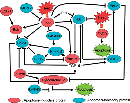
Figure 4. A proposed model for a heat-induced signal transduction pathway based on data obtained from protein microarray analysis.