443
Views
5
CrossRef citations to date
0
Altmetric
Original

Measuring urethral tissue heat injury temperature of healthy male rabbits during interstitial radiofrequency ablation

, , , , &
Pages 56-64 | Received 30 Aug 2007, Accepted 14 Sep 2008, Published online: 09 Jul 2009

Figures & data

Figure 1. The electrode () was advanced into the penial parenchyma along its long axis. The real-time temperature monitoring thermocouple sensor (- - -) was placed parallel into the urethra (△). Correspondence of the tip of needle to the head of thermocouple sensor was assured.
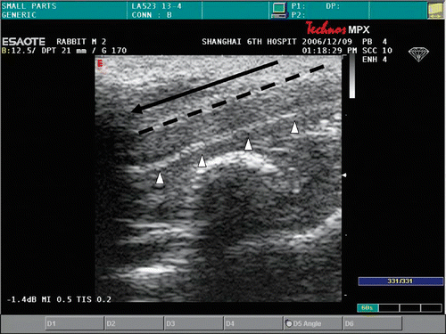
Figure 1. The electrode (–) was advanced into the penial parenchyma along its long axis. The real-time temperature monitoring thermocouple sensor (- - -) was placed parallel into the urethra (△). Correspondence of the tip of needle to the head of thermocouple sensor was assured.

Table I.  Measured parameters of rabbits during experiment.

Figure 2. The histological section showed the urethral mucosa and submucosa tissue surrounding the urethra were displayed clearly in control tissue (▴). The intact cell is characterized by round to oval nuclei with delicately stippled chromatin, preserved spindled cell shape without cytoplasmic retraction, and pale eosinophilic cytoplasm (↑).
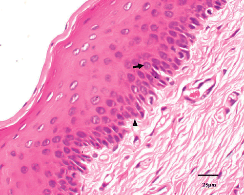
Figure 2. The histological section showed the urethral mucosa and submucosa tissue surrounding the urethra were displayed clearly in control tissue (▴). The intact cell is characterized by round to oval nuclei with delicately stippled chromatin, preserved spindled cell shape without cytoplasmic retraction, and pale eosinophilic cytoplasm (↑).

Figure 3. Coagulative necrosis is characterized by indistinct cellular detail with relatively preserved tissue structure, including pyknotic to karyorectic nuclei and shrunken hypereosinophilic cytoplasm (↑).
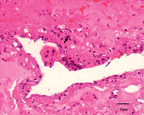
Figure 3. Coagulative necrosis is characterized by indistinct cellular detail with relatively preserved tissue structure, including pyknotic to karyorectic nuclei and shrunken hypereosinophilic cytoplasm (↑).

Figure 4. Mean necrosis percentages measured histologically in control (N = 1), sham-treated (N = 6) and RF-treated groups (N = 5/group). Also shown are results from untreated control animal (N = 1) for comparison. **P < 0.01.
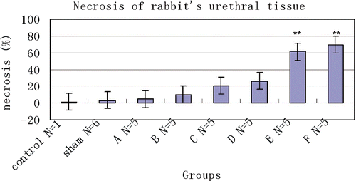
Figure 4. Mean necrosis percentages measured histologically in control (N = 1), sham-treated (N = 6) and RF-treated groups (N = 5/group). Also shown are results from untreated control animal (N = 1) for comparison. **P < 0.01.

Table II.  Necrosis and IOD of urethral tissue with different temperature after RFA.

Figure 5. TUNEL staining on the samples demonstrated absence of apoptotic cells in the normal tissue. The intact cell is characterized by round to oval nuclei without TUNEL-stained chromatin (↑).
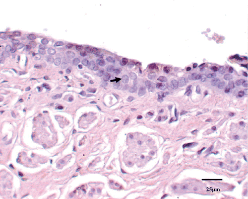
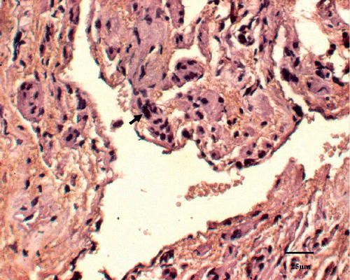
Figure 5. TUNEL staining on the samples demonstrated absence of apoptotic cells in the normal tissue. The intact cell is characterized by round to oval nuclei without TUNEL-stained chromatin (↑).
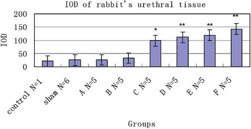
Figure 6. Dead cells by apoptosis stain were identified as those with a red-brown staining of the nucleus indicating the presence of nuclear DNA fragmentation (↑).

Figure 6. Dead cells by apoptosis stain were identified as those with a red-brown staining of the nucleus indicating the presence of nuclear DNA fragmentation (↑).

Figure 7. Mean IOD of TUNEL stain tissue in control (N = 1), sham-treated (N = 6) and RF-treated groups (N = 5/group). Also shown are results from the untreated control animal (N = 1) for comparison. *P < 0.05, **P < 0.01.

Figure 7. Mean IOD of TUNEL stain tissue in control (N = 1), sham-treated (N = 6) and RF-treated groups (N = 5/group). Also shown are results from the untreated control animal (N = 1) for comparison. *P < 0.05, **P < 0.01.

Reprints and Corporate Permissions

Please note: Selecting permissions does not provide access to the full text of the article, please see our help page How do I view content?

To request a reprint or corporate permissions for this article, please click on the relevant link below:

Academic Permissions

Please note: Selecting permissions does not provide access to the full text of the article, please see our help page How do I view content?

Obtain permissions instantly via Rightslink by clicking on the button below:

If you are unable to obtain permissions via Rightslink, please complete and submit this Permissions form. For more information, please visit our Permissions help page.