572
Views
19
CrossRef citations to date
0
Altmetric
Original

The efficacy and safety of percutaneous microwave coagulation by a new microwave delivery system in large hepatocellular carcinomas: Four case studies

, , , , &
Pages 392-398 | Received 02 Oct 2008, Accepted 16 Apr 2009, Published online: 09 Sep 2009

Figures & data

Figure 1. The internally cooled microwave electrode of Forsea Microwave™.

Figure 1. The internally cooled microwave electrode of Forsea Microwave™.

Figure 2. Transverse contrast-enhanced CT scans in patient 1. (a) Pre-ablation scan showed a 6.1 × 5.2 cm cancer nodule (indicated by the wide arrow) in the right lobe. (b) Scan obtained two weeks after one session of ablation showed a complete ablation with a 4.9 × 4.3-cm coagulation zone (indicated by the wide arrow).
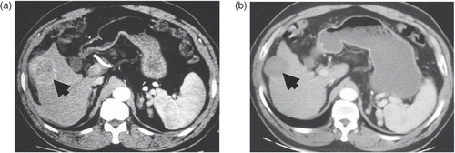
Figure 2. Transverse contrast-enhanced CT scans in patient 1. (a) Pre-ablation scan showed a 6.1 × 5.2 cm cancer nodule (indicated by the wide arrow) in the right lobe. (b) Scan obtained two weeks after one session of ablation showed a complete ablation with a 4.9 × 4.3-cm coagulation zone (indicated by the wide arrow).

Figure 3. Transverse contrast-enhanced CT scans in patient 2. (a) Pre-ablation scan showed a 6.4 × 6.1 cm cancer nodule (arrows) in the right lobe. (b) Scan obtained one month after the first session of ablation showed an incomplete ablation (residual area indicated by the narrow arrow) with a 6.4 × 5.3-cm coagulation zone (indicated by the wide arrow). (c) Scan obtained two weeks after the second session of ablation showed a complete ablation with a 5.8 × 5.2-cm coagulation zone (indicated by the wide arrow).
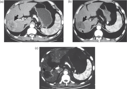
Figure 3. Transverse contrast-enhanced CT scans in patient 2. (a) Pre-ablation scan showed a 6.4 × 6.1 cm cancer nodule (arrows) in the right lobe. (b) Scan obtained one month after the first session of ablation showed an incomplete ablation (residual area indicated by the narrow arrow) with a 6.4 × 5.3-cm coagulation zone (indicated by the wide arrow). (c) Scan obtained two weeks after the second session of ablation showed a complete ablation with a 5.8 × 5.2-cm coagulation zone (indicated by the wide arrow).

Figure 4. Transverse contrast-enhanced CT scans in patient 3. (a) Pre-ablation scan showed a 13.8 × 9.2-cm cancer nodule (indicated by the wide arrow). (b) Scan obtained two weeks after the first session of ablation showed an incomplete ablation (residual area indicated by the narrow arrow) with a 10 × 6.5-cm coagulation zone (indicated by the wide arrow). (c) Scan obtained two weeks after the second session of ablation showed a complete ablation (except the PVTT lesion) with a 7.3 × 6.1 cm coagulation zone (indicated by the wide arrow).
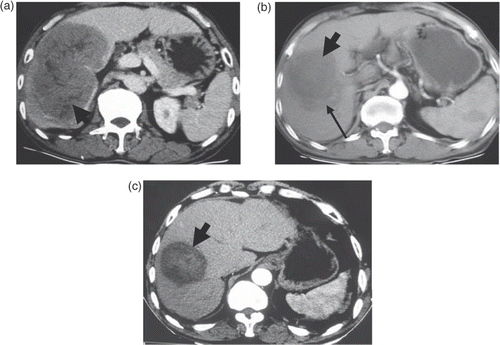
Figure 4. Transverse contrast-enhanced CT scans in patient 3. (a) Pre-ablation scan showed a 13.8 × 9.2-cm cancer nodule (indicated by the wide arrow). (b) Scan obtained two weeks after the first session of ablation showed an incomplete ablation (residual area indicated by the narrow arrow) with a 10 × 6.5-cm coagulation zone (indicated by the wide arrow). (c) Scan obtained two weeks after the second session of ablation showed a complete ablation (except the PVTT lesion) with a 7.3 × 6.1 cm coagulation zone (indicated by the wide arrow).

Figure 5. Transverse contrast-enhanced CT scans in patient 4. (a) Pre-ablation scan showed a 9.8 × 5.2-cm cancer nodule (indicated by the wide arrow). (b) Scan obtained one week after the first session of ablation showed an incomplete ablation (residual area indicated by the narrow arrow) with a 8.9 × 4.8-cm coagulation zone (indicated by the wide arrow). (c) Scan obtained two weeks after the second session of ablation showed an incomplete ablation (residual area indicated by the narrow arrow) with a 8.3 × 5.2-cm coagulation zone (indicated by the wide arrow). (d) Scan obtained two weeks after the third session of ablation showed a complete ablation with a 7.4 × 4.8-cm coagulation zone (indicated by the wide arrow).
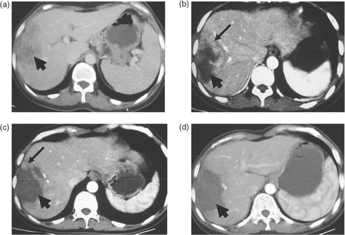
Figure 5. Transverse contrast-enhanced CT scans in patient 4. (a) Pre-ablation scan showed a 9.8 × 5.2-cm cancer nodule (indicated by the wide arrow). (b) Scan obtained one week after the first session of ablation showed an incomplete ablation (residual area indicated by the narrow arrow) with a 8.9 × 4.8-cm coagulation zone (indicated by the wide arrow). (c) Scan obtained two weeks after the second session of ablation showed an incomplete ablation (residual area indicated by the narrow arrow) with a 8.3 × 5.2-cm coagulation zone (indicated by the wide arrow). (d) Scan obtained two weeks after the third session of ablation showed a complete ablation with a 7.4 × 4.8-cm coagulation zone (indicated by the wide arrow).

Reprints and Corporate Permissions

Please note: Selecting permissions does not provide access to the full text of the article, please see our help page How do I view content?

To request a reprint or corporate permissions for this article, please click on the relevant link below:

Academic Permissions

Please note: Selecting permissions does not provide access to the full text of the article, please see our help page How do I view content?

Obtain permissions instantly via Rightslink by clicking on the button below:

If you are unable to obtain permissions via Rightslink, please complete and submit this Permissions form. For more information, please visit our Permissions help page.