2,254
Views
20
CrossRef citations to date
0
Altmetric
Reviews

Status and advancement of microwave ablation in China

&
Pages 278-287 | Received 31 Jul 2016, Accepted 27 Sep 2016, Published online: 20 Dec 2016

Figures & data

Table 1. Commercial MWA system and antenna in China currently.

Table 2. Major researches of MWA in primary and secondary liver malignancies treatment.

Table 3. Major researches of MWA in renal tumours treatment.

Table 4. Results of Chinese clinical studies regarding MWA of lung tumours.
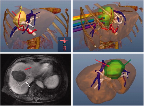
Figure 1. Images of a 70-year-old female patient with a tumour at the border of the left and right lobe. (A) Preoperative 3D images visualised the spatial relationship of the tumour (red arrow) and the surrounding nutrient artery (yellow arrow), portal vein (blue arrow) and hepatic vein (white arrow) in multiple angles. (B) Ablation protocol was achieved through three-dimensional visualisation of the preoperative planning system, and six needles were needed to ablate the tumour completely. (C) The contrast-enhanced magnetic resonance imaging showed complete tumour necrosis (red arrow) one month after microwave ablation without hepatic vein (white arrow) injury. (D) Three-dimensional visualisation software system showed that the ablation zone (green arrow) covered the tumour (red arrow) completely one month after microwave ablation.

Figure 1. Images of a 70-year-old female patient with a tumour at the border of the left and right lobe. (A) Preoperative 3D images visualised the spatial relationship of the tumour (red arrow) and the surrounding nutrient artery (yellow arrow), portal vein (blue arrow) and hepatic vein (white arrow) in multiple angles. (B) Ablation protocol was achieved through three-dimensional visualisation of the preoperative planning system, and six needles were needed to ablate the tumour completely. (C) The contrast-enhanced magnetic resonance imaging showed complete tumour necrosis (red arrow) one month after microwave ablation without hepatic vein (white arrow) injury. (D) Three-dimensional visualisation software system showed that the ablation zone (green arrow) covered the tumour (red arrow) completely one month after microwave ablation.

Reprints and Corporate Permissions

Please note: Selecting permissions does not provide access to the full text of the article, please see our help page How do I view content?

To request a reprint or corporate permissions for this article, please click on the relevant link below:

Academic Permissions

Please note: Selecting permissions does not provide access to the full text of the article, please see our help page How do I view content?

Obtain permissions instantly via Rightslink by clicking on the button below:

If you are unable to obtain permissions via Rightslink, please complete and submit this Permissions form. For more information, please visit our Permissions help page.