Figures & data
Figure 1. Flow chart of the trial. After ablation was finished, CECT or CEMRI was obtained at one month. *For the 3 Grade B tumours, additional treatment was not obtained due to the high risk to damage the surrounding vital structures (n = 1) or poor liver function reserve (n = 2).
US: ultrasound; RFA: radiofrequency ablation; MWA: microwave ablation; CEUS: contrast-enhanced ultrasound.
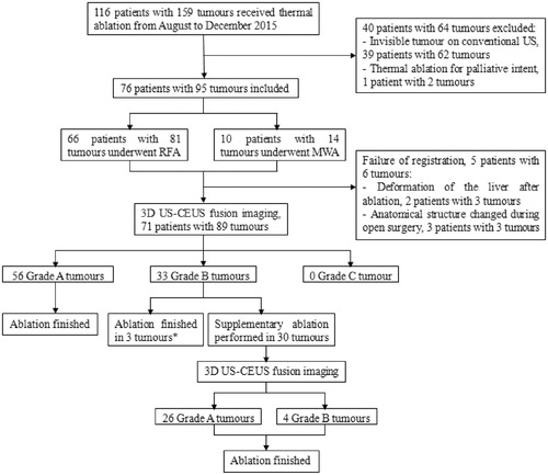
Table 1. Baseline characteristics of the sample.
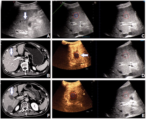
Figure 2. A 82-year-old female patient had a 24 mm HCC tumour in segment 8, which was shown (arrow) on (A) 2D US and (B) CEMRI. (C) The 3D US-US fusion imaging of the lesion before RFA. The real-time US image is shown at left, and the pre-ablation 3D US image is shown at right. (D) Assessment of the ablation effect on 3D US-CEUS fusion imaging after RFA. The non-perfusion area covered the entire lesion with 5 mm AM. The assessment result was Grade A. (E) CEMRI one month after RFA demonstrated the technique effectiveness of the ablation (arrow).
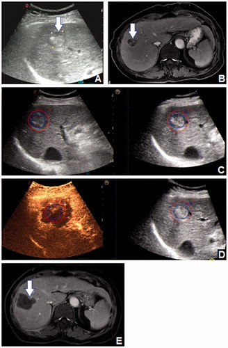
Figure 3. A 61-year-old male patient had a 16 mm HCC tumour in segment 5, which was shown (arrow) on (A) 2D US and (B) CECT. (C) The 3D US-US fusion imaging of the lesion before RFA. The real-time US image is shown at left, and the pre-ablation 3D US image is shown at right. (D) Initial assessment of the ablation effect on 3D US-CEUS fusion imaging after RFA. The non-perfusion area covered the entire lesion, but part of the AM was not covered (arrow). The assessment result was Grade B. (E) Ultimate assessment of the ablation effect on 3D US-CEUS fusion imaging after supplementary ablation. The non-perfusion area covered the entire lesion and the 5 mm AM. The assessment result was Grade A. (F) CECT one month after RFA demonstrated the technique effectiveness of the ablation (arrow).