Figures & data
Table 1. Advantages and disadvantages of the ablative techniques currently used in brain surgery.
Figure 1. A neurosurgeon performing a stereotactic procedure at the hospital “Istituto Neurologico Carlo Besta” in Milan in the early 1980s.
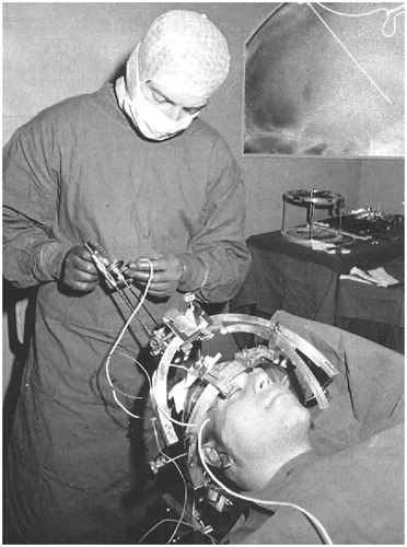
Figure 2. A simulation of a stereotactic MRgFUS ablative procedure with the Kranion software. Temperature prediction is showed on the left side of the panel (maximal: red, average: green). MRgFUS: magnetic resonance-guided focused ultrasound.
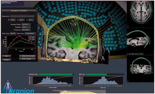
Figure 3. Axial (left) and coronal (right) T2-weighted MRIs demonstrating unilateral thalamotomy (A), pallidotomy (B), and subthalamotomy (C) using transcranial MRgFUS. MRI: magnetic resonance imaging; MRgFUS: magnetic resonance-guided focused ultrasound.
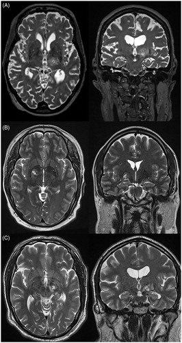
Table 2. Lesion procedures.
Figure 4. Axial T1-weighted MRI sequence demonstrating bilateral anterior capsulotomy using stereotactic radiosurgery for obsessive-compulsive disorder. MRI: magnetic resonance imaging.
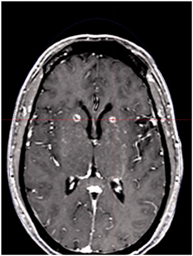
Table 3. Most common brain ablative procedures performed for chronic pain.
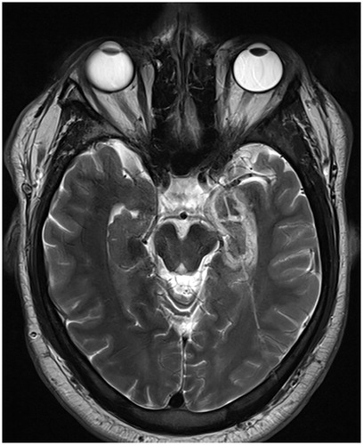
Figure 5. Axial T2-weighted MRI demonstrating left-sided amygdalohippocampectomy and the associated catheter tract following LITT for MTLE. LITT: laser interstitial thermal therapy; MRI: magnetic resonance imaging; MTLE: mesial temporal lobe epilepsy.