Figures & data
Figure 1. R3230 cells survival curves post moderate hyperthermia. In vitro heating of R3230 to 43 °C (A) or 45 °C (B) for 5, 10 or 20 min leads to 80–90% cell survival.
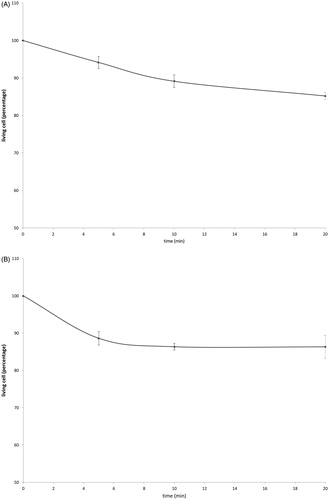
Figure 2. Moderate hyperthermia induces pro-tumorigenic factors mRNA expression in R3230. In vitro heating of R3230 leads to increased expression of cytokines and growth factors. R3230 cells were heated to 43 °C for 5 or 10 min and incubated thereafter at 37 °C for 1.5, 3, and 8 h. The chart presents mRNA fold induction of each labeled cytokine.
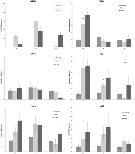
Table 1. mRNA expression levels of studied cytokines following moderate hyperthermia for R3230 and human cancer cell lines.
Figure 3. Moderate hyperthermia induces pro-tumorigenic factors in R3230. (A) In vitro heating of R3230 leads to increase in IL-6 secretion, showed by ELISA. Bar charts indicate a significant increase in IL-6 secretion in medium of R3230 cells that were heated to 43 °C for 5 min followed by incubation of 3 h or 8 h at 37 °C. (B) Western blot assay show an increase in phosphorylation of STAT3 following moderate hyperthermia (dense bands on gel electrophoresis at 80-kDa level as expected, after β-actin standardization). R3230 cells were heated to 43 °C for 5 or 10 min and incubated thereafter at 37 °C for 3 h. Bar charts indicate a significant increase in STAT3 phosphorylation in cells that were heated for 5 min. (C) Western blot assay show increase in c-Met receptor protein following moderate hyperthermia seen as dense bands on gel electrophoresis at 50-kDa level, where c-Met receptor α subunit is expected, after β-actin standardization. Bar charts indicate a significant increase in c-Met receptor protein in R3230 cells that were heated both 5 and 10 min at 43 °C and incubated thereafter at 37 °C for 8 h.
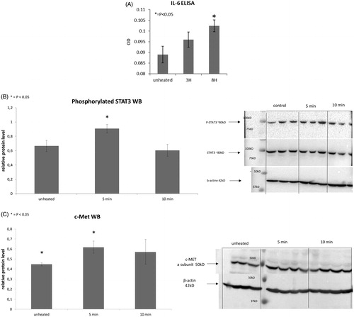
Figure 4. R3230 cells subject to moderate hyperthermia can promote their own accelerated cell growth. Medium of R3230 cells heated at 43 °C for 5 min followed by 8 h incubation was added to naïve R3230 cell culture and enhanced growth of unheated cells compared to the control (C) medium-unheated condition medium. (‘cell index’ refers to confluency of the well.).
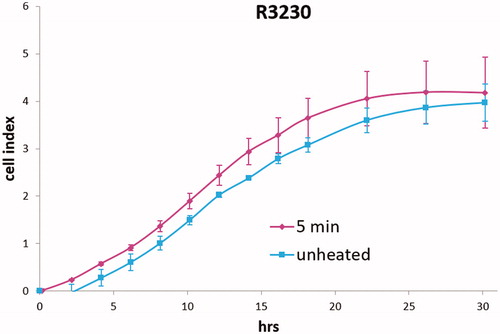
Figure 5. Cancer cells subject to moderate hyperthermia can promote their own accelerated cell growth. Medium of human cancer cells heated at 45 °C for 5 min or 10 min followed by 3 h incubation was added to naïve cell culture and enhanced growth of naïve unheated cells compared to unheated media. Media from human breast MCF7 cells (A), human hepatocellular carcinoma Huh7 (B) and HepG2 cells (C), and human colorectal HT-29 (D) and cells SW480 (E).
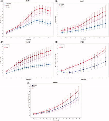
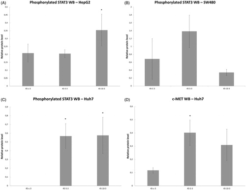
Figure 6. Moderate hyperthermia induces pro-tumorigenic factors in Human cell lines. Western blot assays show an increase in phosphorylation of STAT3 and c-MET receptor α subunit following moderate hyperthermia. cells were heated to 45 °C for 5 or 10 min and incubated thereafter at 37 °C for 3 h. Bar charts indicate an increase in STAT3 phosphorylation or c-MET in cells that were heated. (A) HepG2 cells show an increase in pSTAT3 10 min post heating. (B) SW480 cells show an increase in pSTAT3 5 min post heating. (C) Huh7 cells show an increase in pSTAT3 5 and 10 min post heating. (D) Huh7 cells show an increase in c-MET receptor α subunit 5 and 10 min post heating.