4,661
Views
59
CrossRef citations to date
0
Altmetric
Reviews

In vivo magnetic nanoparticle hyperthermia: a review on preclinical studies, low-field nano-heaters, noninvasive thermometry and computer simulations for treatment planning

ORCID Icon, & ORCID Icon
Pages 76-99 | Received 15 Apr 2020, Accepted 17 Jul 2020, Published online: 10 Jan 2021

Figures & data

Table 1. Potential thermometry techniques compatible with in vivo MNH, limitations and advantages.

Table 2. Preclinical studies that show the effect of MNH (alone) in the treatment of solid tumors.

Figure 1. In several preclinical studies reported on the literature, the experimental setups for in vivo MNH are out of the clinical safe-limit region (some much more than others): at the graph the dashed and shaded areas delimits, respectively, the Atkinson1 and Dutz-Hergt2 thresholds to prevent harmful non-localized heating regime due to eddy currents. The points correspond to the ordered pair of the experimental values for frequency f (in kHz) and amplitude H (in kA.m−1 and Oe) of the AMF for the reports presented in .

Figure 1. In several preclinical studies reported on the literature, the experimental setups for in vivo MNH are out of the clinical safe-limit region (some much more than others): at the graph the dashed and shaded areas delimits, respectively, the Atkinson1 and Dutz-Hergt2 thresholds to prevent harmful non-localized heating regime due to eddy currents. The points correspond to the ordered pair of the experimental values for frequency f (in kHz) and amplitude H (in kA.m−1 and Oe) of the AMF for the reports presented in Table 2.

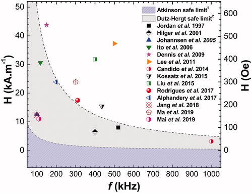
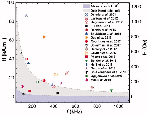
Table 3. Some mono-core and multicore nano-heaters (from 2008 to 2019), targeted for better SLP performance with in vivo MNH.

Figure 2. In several in vitro studies reported on the literature, the experimental MNH setups are out of the clinical safe-limit region (some much more than others): in the graph the dashed and shaded areas delimit, respectively, the Atkinson1 and Dutz-Hergt2 thresholds to prevent harm in human applications (minimizing the off-target heating due to eddy currents). The points correspond to the ordered pair of the experimental values for frequency f (in kHz) and amplitude H (in kA.m−1 and Oe) of the AMF for each work presented in .

Figure 2. In several in vitro studies reported on the literature, the experimental MNH setups are out of the clinical safe-limit region (some much more than others): in the graph the dashed and shaded areas delimit, respectively, the Atkinson1 and Dutz-Hergt2 thresholds to prevent harm in human applications (minimizing the off-target heating due to eddy currents). The points correspond to the ordered pair of the experimental values for frequency f (in kHz) and amplitude H (in kA.m−1 and Oe) of the AMF for each work presented in Table 3.

Table 4. Computer simulations for in vivo MNH.

Figure 3. In vivo MNH treatment planning algorithm. MRI – Magnetic Resonance Imaging, CT – Computed Tomography, US – Ultrasound, US-SW – Shear wave ultrasound, MPI – Magnetic Particle Imaging, FMT - Fluorescence Molecular Tomography, * Technique useful only for preclinical studies.

Figure 3. In vivo MNH treatment planning algorithm. MRI – Magnetic Resonance Imaging, CT – Computed Tomography, US – Ultrasound, US-SW – Shear wave ultrasound, MPI – Magnetic Particle Imaging, FMT - Fluorescence Molecular Tomography, * Technique useful only for preclinical studies.