
Figures & data
Figure 1. High-speed photograph of cavitation generated by (A) intrinsic threshold histotripsy (modified from ref [Citation69]) and (B) shockscattering histotripsy (modified from ref [Citation73]). Ultrasound propagates from left to right.
![Figure 1. High-speed photograph of cavitation generated by (A) intrinsic threshold histotripsy (modified from ref [Citation69]) and (B) shockscattering histotripsy (modified from ref [Citation73]). Ultrasound propagates from left to right.](/cms/asset/b8274990-da4e-4455-a454-aa30bf5b9577/ihyt_a_1905189_f0001_b.jpg)
Figure 2. (A) H&E slide of an ex vivo porcine cardiac tissue treated by histotripsy shows no intact cells within the treated region and bisected cells at the sharp lesion boundary. (B–C) Gross morphology of the rat liver treated by histotripsy shows a darkly colored hematoma within the histotripsy-treated zone right after treatment (B) and only a small contracted scar on Day 28 (C). (D) Histology of the rat liver treated by histotripsy shows a small focus of residual scar and foreign body giant cells (GC) with calcified undigested debris on Day 28. (B–D) are reproduced based on images from ref [Citation22]. (E) A perforation is generated by histotripsy through a piece of ex vivo porcine atrial wall.
![Figure 2. (A) H&E slide of an ex vivo porcine cardiac tissue treated by histotripsy shows no intact cells within the treated region and bisected cells at the sharp lesion boundary. (B–C) Gross morphology of the rat liver treated by histotripsy shows a darkly colored hematoma within the histotripsy-treated zone right after treatment (B) and only a small contracted scar on Day 28 (C). (D) Histology of the rat liver treated by histotripsy shows a small focus of residual scar and foreign body giant cells (GC) with calcified undigested debris on Day 28. (B–D) are reproduced based on images from ref [Citation22]. (E) A perforation is generated by histotripsy through a piece of ex vivo porcine atrial wall.](/cms/asset/760161ae-8e89-42bf-8ef8-f15595eb3f8a/ihyt_a_1905189_f0002_c.jpg)
Figure 3. (A) Large blood vessels remain intact and surrounded by acellular debris within the in vivo porcine liver treated by histotripsy (reproduced based on an image from ref [Citation37]. (B) Contrast-enhanced MR image showing a patent bile duct (blue arrow) within the dark histotripsy treatment zone (reproduced based on an image from ref [Citation84].
![Figure 3. (A) Large blood vessels remain intact and surrounded by acellular debris within the in vivo porcine liver treated by histotripsy (reproduced based on an image from ref [Citation37]. (B) Contrast-enhanced MR image showing a patent bile duct (blue arrow) within the dark histotripsy treatment zone (reproduced based on an image from ref [Citation84].](/cms/asset/81d51ffe-f58b-470d-bd9a-9ada45bb3e13/ihyt_a_1905189_f0003_c.jpg)
Table 1. Typical histotripsy parameters.
Figure 4. An ultrasound-guided histotripsy system that contains an ultrasound imaging engine, a histotripsy transducer with the imaging probe in the center (insert), a positioning arm that mechanically moves the histotripsy transducer.
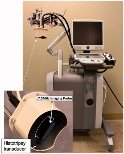
Figure 5. (A) Histotripsy-generated cavitation is seen as a hyperechoic zone on B-mode ultrasound. (B) ‘M’ shaped histotripsy lesion shows as a hypoechoic zone on B-mode ultrasound image. (C) Histotripsy ablation zone is seen as a hypointense (non-enhancing) region on this T1-weighted contrast-enhanced MR image.
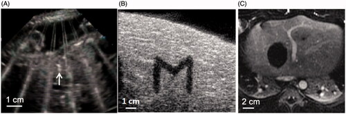
Figure 6. (A–D) Histotripsy treatment in the in vivo porcine liver. Axial T2-weighted MR images (A) and gross morphology (B) show the spherical ablation volume. (C) H&E slide of (100X) of the center of the ablation zone demonstrates no viable hepatocytes. (D) H&E slide (100X) of the periphery of the ablation shows viable liver (L), necrotic liver (N), and a thin zone of transition (Z). (E–G) MR images of partial histotripsy treatment in a rodent HCC model. (E) The appearance of the original untreated tumor on T2-weighted MRI was hyperintense (yellow arrow). (F) Post histotripsy, the ablation zone demonstrates T2 hypointense signal (red dashed lines). (G) At 12 weeks, only a ∼5 mm non-tumoral fibrous tissue zone is detectable on MRI at the original tumor site, indicating near-complete resolution of the ablation zone and tumor. (H–I) Histotripsy induced immune response and abscopal effect (reproduction based on images from ref [Citation39]. (H) FACS analysis of histotripsy-ablated (HT ablated) and contralateral non-ablated tumors (HT abscopal) identified comparable levels of intratumoral CD8+ T cell infiltration that is significantly higher than the untreated control tumors. (I) At gross examination, pulmonary metastases were reduced in mice treated with histotripsy compared to untreated controls.
![Figure 6. (A–D) Histotripsy treatment in the in vivo porcine liver. Axial T2-weighted MR images (A) and gross morphology (B) show the spherical ablation volume. (C) H&E slide of (100X) of the center of the ablation zone demonstrates no viable hepatocytes. (D) H&E slide (100X) of the periphery of the ablation shows viable liver (L), necrotic liver (N), and a thin zone of transition (Z). (E–G) MR images of partial histotripsy treatment in a rodent HCC model. (E) The appearance of the original untreated tumor on T2-weighted MRI was hyperintense (yellow arrow). (F) Post histotripsy, the ablation zone demonstrates T2 hypointense signal (red dashed lines). (G) At 12 weeks, only a ∼5 mm non-tumoral fibrous tissue zone is detectable on MRI at the original tumor site, indicating near-complete resolution of the ablation zone and tumor. (H–I) Histotripsy induced immune response and abscopal effect (reproduction based on images from ref [Citation39]. (H) FACS analysis of histotripsy-ablated (HT ablated) and contralateral non-ablated tumors (HT abscopal) identified comparable levels of intratumoral CD8+ T cell infiltration that is significantly higher than the untreated control tumors. (I) At gross examination, pulmonary metastases were reduced in mice treated with histotripsy compared to untreated controls.](/cms/asset/5d873b5e-d0a1-4097-908a-98658e74ee3e/ihyt_a_1905189_f0006_c.jpg)