394
Views
1
CrossRef citations to date
0
Altmetric
Research Article

Interaction of 3-hydroxybenzoate-6-hydroxylase with cibacron blue

&
Pages 47-53 | Received 10 Apr 2005, Accepted 10 Aug 2005, Published online: 04 Oct 2008

Figures & data

Figure 1 Effect of cibacron blue on the initial velocities of 3-HBA-6-hydroxylase; a) activity measured by fluorimetric assay. b) activity measured by polarographic method.
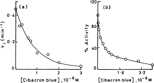
Figure 1 Effect of cibacron blue on the initial velocities of 3-HBA-6-hydroxylase; a) activity measured by fluorimetric assay. b) activity measured by polarographic method.

Figure 2 Double reciprocal plots for demonstrating the nature of inhibition of 3-HBA-6-hydroxylase activity by cibacron blue with respect to 3-HBA.
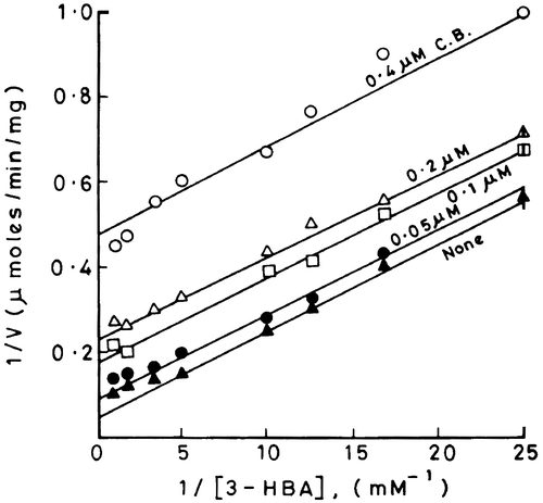
Figure 2 Double reciprocal plots for demonstrating the nature of inhibition of 3-HBA-6-hydroxylase activity by cibacron blue with respect to 3-HBA.

Figure 3 Profile of quenching of intrinsic fluorescence (emission maximum at 335 nm) of 3-HBA-6- hydroxylase by cibacron blue.
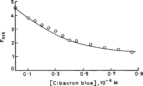
Figure 3 Profile of quenching of intrinsic fluorescence (emission maximum at 335 nm) of 3-HBA-6- hydroxylase by cibacron blue.

Figure 4 Double reciprocal plots for the observed changes in enzyme fluorescence with increasing concentrations of cibacron blue using native enzyme, FAD-enzyme, 3-HBA-enzyme and urea denatured enzyme.
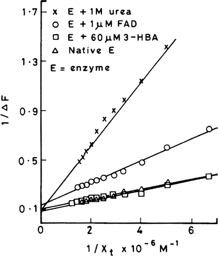
Figure 4 Double reciprocal plots for the observed changes in enzyme fluorescence with increasing concentrations of cibacron blue using native enzyme, FAD-enzyme, 3-HBA-enzyme and urea denatured enzyme.

Figure 5 Stinson and Holbrook plots for the quenching of enzyme fluorescence with increasing concentrations of cibacron blue using native enzyme, 3-HBA-enzyme, FAD-enzyme and urea denatured enzyme.
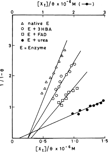
Figure 5 Stinson and Holbrook plots for the quenching of enzyme fluorescence with increasing concentrations of cibacron blue using native enzyme, 3-HBA-enzyme, FAD-enzyme and urea denatured enzyme.

Table I.  Binding parameters, stoichiometry (r) and association constant (Ka), for the enzyme–cibacron blue interaction. The binding parameters were calculated from the equation: 1/(1−θ) Ka=[XT]/θ−r[ET].
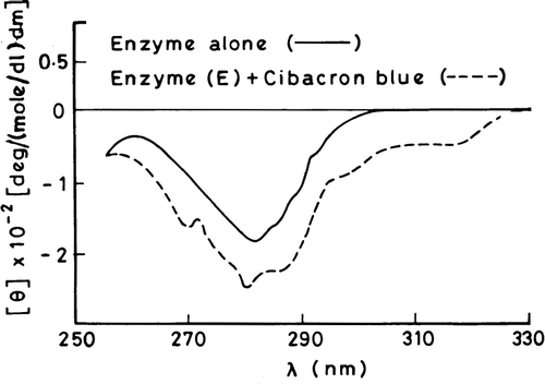
Figure 6 Near-UV CD spectral profiles of 3-HBA-6-hydroxylase in the absence and presence of cibacron blue.

Figure 6 Near-UV CD spectral profiles of 3-HBA-6-hydroxylase in the absence and presence of cibacron blue.

Reprints and Corporate Permissions

Please note: Selecting permissions does not provide access to the full text of the article, please see our help page How do I view content?

To request a reprint or corporate permissions for this article, please click on the relevant link below:

Academic Permissions

Please note: Selecting permissions does not provide access to the full text of the article, please see our help page How do I view content?

Obtain permissions instantly via Rightslink by clicking on the button below:

If you are unable to obtain permissions via Rightslink, please complete and submit this Permissions form. For more information, please visit our Permissions help page.