Figures & data
Table I. QOL analysis in patients with germinomas who received the whole brain radiotherapy between 1976 and 1991.
Table II. Characteristics of patients with germinoma or germinoma STGC since 1992 (total 30 cases).
Table III. Germinoma Recurrence rate in the treatment of each Radiotherapy.
Figure 1. Case 1, a 12-year-old boy. a: Pre-treatment MRI scan revealing a pineal mass. b: Post-treatment MRI scan revealing disappearance of the pineal mass. c: On this x-radiograph, the field of irradiation (gray area) and ventricles (white area) on MRI scans are traced. The relapse site is identified by a hatched circle. d: This post-treatment MRI scan shows an enhanced lesion in the posterior horn of the left lateral ventricle (arrow).
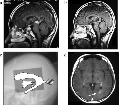
Figure 2. Case 2, a 15-year-old boy. a: Pre-treatment MRI scan revealing a pineal mass. b: Post-treatment MRI scan reveals complete disappearance of the pineal mass. c: On this x-radiograph, the field of irradiation (gray area) and the ventricles (white area) on MRI scans are traced. The relapse site is identified by a hatched circle. d: This post-treatment MRI scan reveals an enhanced lesion in the anterior horn of the right lateral ventricle (arrow).
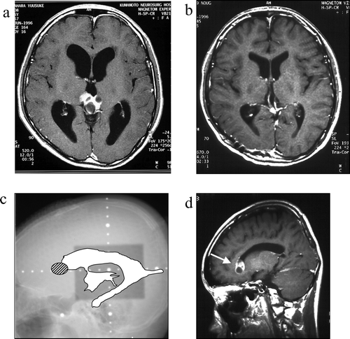
Figure 3. Case 3, a 16-year-old boy. a: Pre-treatment MRI scan revealing an enhanced lesion in the right basal ganglia. b: Post-treatment MRI scan shows complete disappearance of the lesion. c: On this x-radiograph, the field of irradiation (gray area) and the ventricles (white area) on MRI scans are traced. The relapse site is identified by a hatched circle. d: This post-treatment MRI scan (axial view) reveals an enhanced lesion in the inferior horn of the right lateral ventricle (arrow). e: MRI scan obtained at the time of tumor recurrence (coronal view).
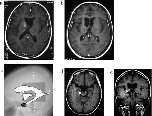
Figure 4. Case 4, an 11-year-old boy. a: Pre-treatment MRI scan revealing a hypothalamic enhanced lesion. b: Post-treatment MRI scan shows complete disappearance of the hypothalamic mass. c: On this x-radiograph, the field of irradiation (gray area) and the ventricles (white area) on MRI scans are traced. The relapse site is identified by a hatched circle. d: MRI scan obtained at the time of tumor recurrence shows an enhanced lesion in the body of the right lateral ventricle (arrow).
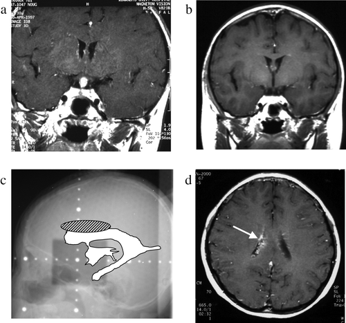
Table IV. Characteristics of the patients with recurrence of the germinoma at the outside of the initial radiation field.