Figures & data
Figure 1. Effects of time held in suspension on spheroid volume. Relative volumes of spheroids composed exclusively of live (black squares), and supralethally irradiated (20 Gy, open squares) AG1522 fibroblasts are plotted for successive days after spheroid selection. Both types of spheroids shrank to about 10–20% of their original size within three days (day four after creation), after which, they remain the same reduced size for the rest of the period of observation (up to two weeks). In contrast, pure spheroids of HeLa cells (black circles) grow, increasing in size (volume) by an order of magnitude over a 6-day period. Data obtained from four experiments. Error bars in this and in other graphs are standard errors of the mean.
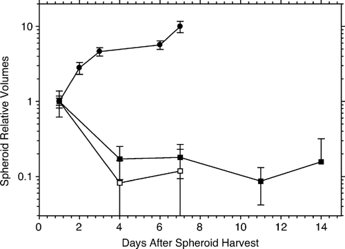
Figure 2. Increases in the relative number of clonogens with time in culture. The top two curves with solid lines are for HeLa cells in either attached (open squares) or floating (open circles) pure spheroids. For comparison, a growth curve of HeLa cells in monolayer is also shown (open stars, dashed line). The next two curves represent the growth of HeLa cells in hybrid spheroids (with irradiated AG1522 fibroblast feeder cells; open triangles) and of unirradiated AG1522 fibroblasts grown in monolayer (black stars). The bottom curve (black circles) shows the initial stasis, and then growth, of AG1522 fibroblasts as test cells in attached hybrid spheroids with irradiated HeLa feeder cells; the final growth seen between days 3 and 4 is similar to that of the HeLa cells in floating or attached pure spheroids or in monolayer.
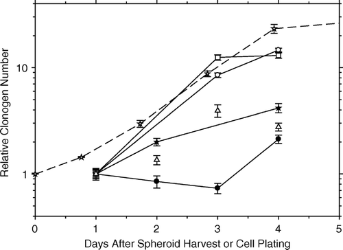
Figure 3. The loss of clonogenic AG1522 fibroblasts maintained in spheroids. The bottom two curves show the rapid loss of clonogenic fibroblasts in: (1) floating hybrid spheroids with irradiated HeLa feeder cells (black circles) and (2) in floating pure spheroids (black squares). Within three days after harvest, only about 1–2 percent of fibroblasts remain clonogenic; within four days < 3 per 1 000 remain clonogenic. In both cases, the spheroids were trypsinized before plating for colony formation. The top curve (black triangles) shows the loss of clonogenicity with time for AG1522 fibroblasts in pseudohybrid spheroids trypsinized prior to plating for colonies.
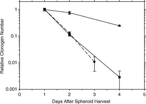
Figure 4. The slow loss of clonogenicity of AG1522 fibroblasts in pure and in pseudohybrid spheroids left intact for plating for colonies to grow out of each marked spheroid, i.e., NOT trypsinized. The top two curves show the results for pure spheroids. One curve is for attached spheroids, the positions of which had been marked, and clonogenicity was calculated from the fraction which did not form a colony (open square). The other is for floating pure spheroids (black square), which were plated at the indicated time and later scored for how many colonies they had produced. The lower two curves (solid lines) are for pseudohybrid fibroblast spheroids. The upper one (open circles) is for floating spheroids (as above), while the lower one (black circles) is for attached spheroids (as above). The curve for trypsinized floating pseudohybrid fibroblast spheroids is shown for comparison (from , black triangles, dashed line), and is essentially the same as that for attached pseudohybrid spheroids.
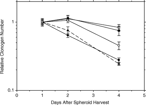
Figure 5. Survival curves of HeLa cells irradiated in hybrid spheroids, corrected for clonogen multiplicity. Hybrid spheroids were maintained in suspension over a period of 10–12 days before scoring surviving spheroids. Data obtained from four experiments in each series. Black circles: live fibroblast feeder cells, Do=2.95±0.08 Gy (α = 0.339±0.009 Gy−1). White circles: supralethally (20 Gy) irradiated fibroblast feeder cells, Do=1.92±0.15 Gy (α = 0.52±0.04 Gy−1). The ratio of D0 values = 1.54±0.13.
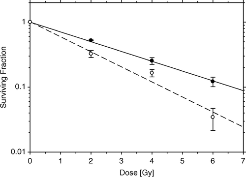
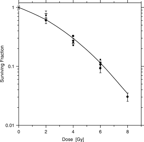
Figure 6. Fibroblast feeder cells (dead or alive) do not affect the radiosensitivity of HeLa test cells in spheroids dispersed before irradiation. HeLa cell survival curves were obtained for cells dispersed, irradiated and then incubated for 10–12 days before scoring colonies and fitting the Linear-Quadratic equation to the data. Dispersed monolayer cultures with (squares) and without (circles) irradiated feeder cells, dispersed hybrid spheroids containing live fibroblast feeders (triangles), or dispersed hybrid spheroids containing supralethally irradiated fibroblast feeders (inverted triangles). Parameters from common curve, α = 0.18±0.03 Gy−1, β = 0.030±0.008 Gy−2.