Figures & data
Figure 1. Diagram demonstrating the role of E-Cadherin; histologically, cellularly and molecularly in EMT. E-Cadherin, found at adherens junctions, maintains epithelial cell-cell contact, due to homophilic binding of its extracellular domains. During EMT, apical-basal polarity is lost, E-Cadherin expression is downregulated and mesenchymal genes are up-regulated. The binding of the transcriptional repressors Slug, Sip1, Snail and Twist to the inhibitory E-boxes of the E-Cadherin promoter terminates E-Cadherin expression.
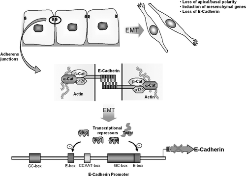
Table I. The importance of EMT and MET in embryogenesis. See text and associated cited references for additional information.
Figure 2. Diagram of the Wnt signaling pathway. (A) In the absence of Wnt ligand, β-catenin undergoes intracellular destruction. (B) In the presence of Wnt ligand, β-catenin accumulates in the cytoplasm and translocates to the nucleus, where it binds to TCF/LEF transcription elements. This effects the transcription of downstream targets.
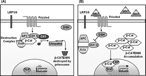
Figure 3. Diagram of the Sonic Hedgehog pathway. Sonic Hedgehog binds to the Ptch receptor, this relieves inhibition of Smoothened. Downstream this causes upregulation of Gli zinc finger transcription factors. SUFU is an intracellular molecule that forms a repressor unit that inhibits Gli activities.
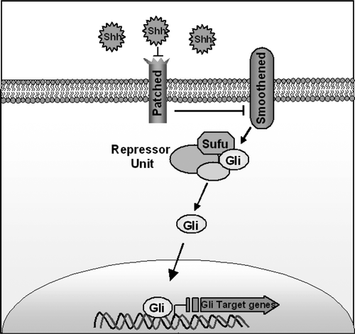
Table II. The role of Sonic Hedgehog, Desert Hedgehog, Indian Hedgehog and Ptch in embryogenesis. See text and associated cited references for additional information.
Figure 4. Diagram of the Notch Pathway: Pathway activation by delta or serrate ligands cleaves the Notch receptor and causes translocation of its intracellular portion (ICN) to the nucleus. ICN forms part of an enhancesome that results in the transcription of target genes.
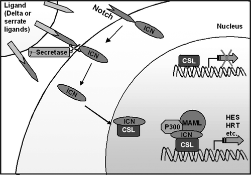
Table III. Alterations in the Notch Pathway in selected malignancies