Figures & data
Figure 1. The radiograph demonstrates an ill defined lytic lesion involving medial two thirds of the clavicle with a soft tissue shadow extending beyond the confines of the bone.
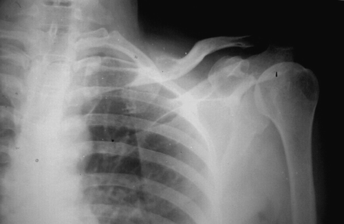
Figure 2. The computed tomographic images of the same patient reveals a ballooned out lesion (1) involving the medial two thirds of the clavicle with a thin shell of bone around. There is extension of the lesion into soft tissue (3) and cortical breach is well demonstrated (2).
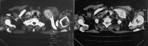
Figure 3. Histopathology: Microscopic examination showed areas with abundant osteoid formation with low degree of cellularity with no atypia. Undecalcified sections depicted partial calcification of osteoid, elsewhere highly cellular areas were present comprising oval osteoblast like cells (epitheloid osteoblasts) and moderate nuclear pleomorphs with occasional mitosis and intermingled tumor giant cells.
