Figures & data
Figure 1. Two examples of individualized PTV generation for SRT of lung cancer using 4DCT scans. GTVs are contoured in each of the 10 phase bins of the 4DCT, and the ITV is defined as the volume encompassing all GTVs. A CTV to PTV margin of 3 mm is added to the ITV to derive the individualized PTV.
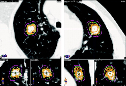
Figure 2. Two-phase planning target volumes based on contouring of the end-inspiration (yellow) and end-expiration (pink) phase bins of the 4DCT for a tumor showing predominantly cranio-caudal mobility.
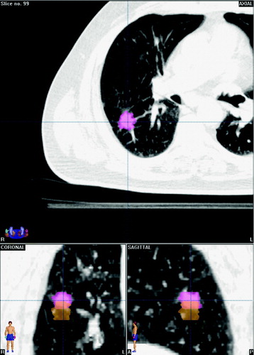
Figure 3. Examples of MIP scans that incorporate tumor mobility from all phases of the 4DCT scan. Left panel: Projection of GTVs from 10 phase bins (yellow) onto a maximum intensity projection (MIP) CT scan, with contouring on the MIP scan shown in red. Right panel: The end-inspiratory (yellow) and end-expiratory (pink) GTVs from the 4DCT of another patient, projected onto the MIP scan.
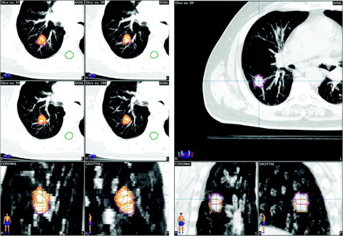
Figure 4. Frontal reconstruction of 4DCT scans in a patient with two tumors in the right lung. Images illustrate the respective maximum intensity projection (a), minimum intensity projection (b), mean intensity projection (c), and color intensity projection (d) protocols for this 4DCT study.
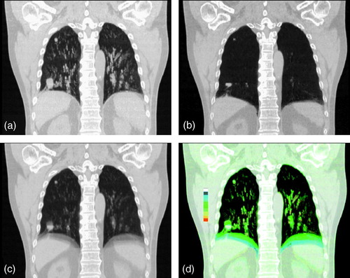
Figure 5. CIP images of a patient with a prior right pneumonectomy, showing mobility of the left diaphragm and left main stem bronchus (left). A tumor in the left lower lobe shows predominantly cranio-caudal mobility (arrow) and the colored ‘column’ indicates the trajectory of mobility. The green color of the most cranial position indicates that the tumor is at its end-expiratory position for 50% of the respiratory cycle.
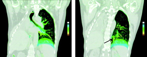
Table I. Stereotactic radiotherapy schemes for Stage I NSCLC tumors at the VUmc