1,370
Views
47
CrossRef citations to date
0
Altmetric
LETTER TO THE EDITOR

Helical tomotherapy targeting total bone marrow – First clinical experience at the University of Minnesota

, , , , , , , & show all
Pages 250-255 | Received 05 Sep 2006, Published online: 08 Jul 2009

Figures & data

Figure 1.  MVCT scan taken at the abdomen before treatment delivery. MVCT scan set was fused with treatment planning CT scan. The transverse sectional views are shown in (A) Tomo MVCT image and in (B) reference kVCT image. The fusion column (C) presents the kVCT in gray scale with the MVCT superimposed with a level of transparency in yellow. Similarly, the sagital sectional views and the coronal sectional views are shown in D, E, F and G, H, I respectively.

Figure 1.  MVCT scan taken at the abdomen before treatment delivery. MVCT scan set was fused with treatment planning CT scan. The transverse sectional views are shown in (A) Tomo MVCT image and in (B) reference kVCT image. The fusion column (C) presents the kVCT in gray scale with the MVCT superimposed with a level of transparency in yellow. Similarly, the sagital sectional views and the coronal sectional views are shown in D, E, F and G, H, I respectively.

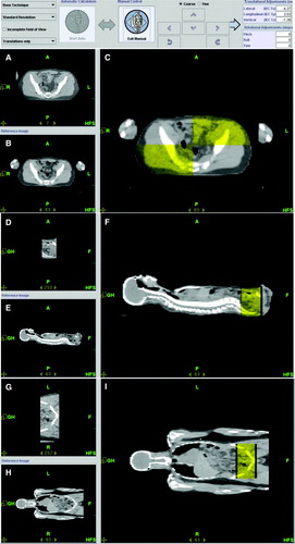
Table I.  Translational (in IEC Tx, Ty, Tz) shift at various anatomical sites for pre-treatment and post-treatment using MVCT-kVCT fusion.

Figure 2.  Sectional view of dose painting: (I) transverse, (II) sagital, (III) coronal view for upper part of the body, and (IV) transverse, (V) sagital, (VI) coronal view for lower part of the body; (VII) The dose volume histogram for PTV(A), liver(B), heart(C), eye(D), lungs(E), spleen(F), left kidney(G), right kidney(H), lens(I), scrotum(J), and (V) DVH for bony anatomy: Upper body PTV (A), thoracic bone(A1); bone of skull(A2); pelvic bone(A3); right extremities(A4) are shown in lower middle panel (VIII). Right lower panel (IX) shows DVH for lower body PTV (A’).

Figure 2.  Sectional view of dose painting: (I) transverse, (II) sagital, (III) coronal view for upper part of the body, and (IV) transverse, (V) sagital, (VI) coronal view for lower part of the body; (VII) The dose volume histogram for PTV(A), liver(B), heart(C), eye(D), lungs(E), spleen(F), left kidney(G), right kidney(H), lens(I), scrotum(J), and (V) DVH for bony anatomy: Upper body PTV (A), thoracic bone(A1); bone of skull(A2); pelvic bone(A3); right extremities(A4) are shown in lower middle panel (VIII). Right lower panel (IX) shows DVH for lower body PTV (A’).

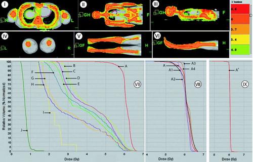
Table II.  Median dose in various organs for prescription dose (Rx) of 6 Gy.

Reprints and Corporate Permissions

Please note: Selecting permissions does not provide access to the full text of the article, please see our help page How do I view content?

To request a reprint or corporate permissions for this article, please click on the relevant link below:

Academic Permissions

Please note: Selecting permissions does not provide access to the full text of the article, please see our help page How do I view content?

Obtain permissions instantly via Rightslink by clicking on the button below:

If you are unable to obtain permissions via Rightslink, please complete and submit this Permissions form. For more information, please visit our Permissions help page.