Figures & data
Figure 1. (A) The expression of TERTt in B16 and CT26 cells. 1: DNA marker I; 2: RT-PCR product of -actin in B16 cells; 3:RT-PCR product of TERTt in B16 cells; 4: RT-PCR product of -actin in CT26 cells; 5: RT-PCR result of CT26 cells using TERTt primers. (B) Linearization of pBluescriptIIKS(+)-TERTt : 1. DNA marker I 2. Linearized pBluescriptIIKS(+)-TERTt template 3.Double enzyme digestion of pBluescriptIIKS(+)-TERTt. (C) In vitro transcription of TERTt mRNA: 1. RNA marker 2.Capped TERTt mRNA 3. Capped TERTt mRNA with poly-A tail.
Figure 2. DCs were eletroporated with EGFP mRNA to evaluate the transfection efficiency. Post EGFP mRNA electroporation 24h, EGFP expression can be detected in CD11c+ DCs using FCM analysis. (A) The DCs of 8th day culture system showed no EGFP expression; (B) After EGFP mRNA electroporated for 24h, The DCs of 7th day culture system showed EGFP expression.
Figure 3. TERTt mRNA transfected DCs immunization can induce B16 specific CTLs in C57B/L mice. (A) TERTt mRNA transfected DC immunization can induce IFN-γ secreting specific Splenocytes responding to Mitomycin C treated B16 cells. 1: Splenocytes of Blank DC immunized mice; 2: Splenocytes of TERTt-DC immunized mice; 3: Splenocytes of hMAGEn-DC immunized mice; 4: Negtive control (Splenocytes of Blank DC immunized mice, no target). 5: Positive control (Splenocytes of Blank DC immunized mice, no target, 1mg/L PHA stimulated). (B) Splenocytes obtained from TERTt mRNA transfected DCs immunized mice showed escalated cytotoxic activity against Mitomycin C treated B16 cells at E:T ratios indicated. ▴: Splenocytes of TERTt-DC immunized mice; •: Splenocytes of Mn-DC immunized mice; ▪: Splenocytes of Blank-DC immunized mice. (Values are the mean±SD of triplicate wells)
Figure 4. (A) Primary CT26 cells showed undetectable TERT activity. After stimulated with target cells for 24 hours, splenocytes derived from TERTt-DC immunized mice only menifested IFN-γ secreting response against CT26-TERTt cells but not CT26 or CT26-hMAGEn cells. 1: Splenocytes of Blank DC immunized mice; 2: Splenocytes of TERTt-DC immunized mice; (Values are the mean±SD of triplicate wells) (B) TERTt mRNA transfected DC immunization can generate TERTt specific CTLs in BALB/c mice in vivo. Splenocytes of TERTt-DC immunized mice displayed strong cytolytic activity against CT26-TERTt at E:T ratio of 10:1, 20:1 and 40:1. Left: Splenocytes of Blank DC immunized mice; Middle: Splenocytes of TERTt-DC immunized mice. (Values are the mean±SD of triplicate wells)
Figure 5. The Survival curve of different mRNA transfected DCs immunized/ B16-challenged mice. Log-rank test showed that the median survival time was different in these three groups of mice (p = 0.029). The mice vaccinated with TERTt-DC showed longer survival time than those vaccinated with Blank DC or hMAGEn-DC. 1: Blank DC immunized mice with B16 challenged; 2: TERTt-DC immunized mice with B16 challenged; 3: hMAGEn-DC immunized mice with B16 challenged.
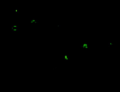
Image1. The TERTt expression can be detected in Dendritic cells by using immunofluorescence assay 24 hours post TERTt mRNA electroporation. (First antibody: rabbit anti mouse TERT; Second antibody: FITC-goat anti rabbit IgG)