Figures & data
Figure 1. Tissue pathology of the biopsied specimen showing a picture of a malignant melanoma with solid nests infiltrating into the mucosa, superficial ulceration, moderate nuclear pleomorphism, and focal pigmentation. Immunohistochemical study revealed that the tumor was positive for HMB-45 and negative for cytokeratin.
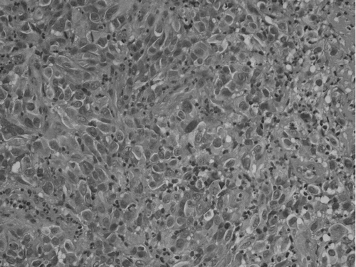
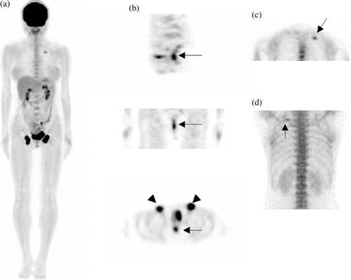
Figure 2. Whole-body PET was performed 45 min after an intravenous injection of 300 MBq FDG using a Siemens ACCEL PET scanner. Additional 3-h-delayed images after emptying the urinary bladder were also obtained. (A) Maximal intensity projection (MIP) imaging revealing focal increases in FDG uptake in the bilateral inguinal regions and in the posterior aspect of the left upper thorax. The rectal region was obscured by the full urinary bladder. (B) Delayed image in the sagittal and coronal sections revealing focal increase activity in the rectum (upper and middle), and a transverse section revealing FDG uptake in the rectum and bilateral inguinal lymph nodes (lower). The maximal standard uptake value (SUVm) of the rectal lesion was 5.9. (C) Coronal section of the thorax revealing a focal area with increase FDG uptake. A corresponding finding was observed in the posterior view of the Tc-99m MDP bone scan which showed a solitary hot spot in the left 4th rib (D).