Figures & data
Figure 1. CT scan of head of patient 1 showing a large recurrent right temporoparietooccipital tumor with mass effect and midline shift.
Figure 2. Photomicrograph of patient 3 showing islands of glial tissue alternating with fibroblastic stroma (a, H&E×100; b H&E×200). Another area showing fibroblastic tissue with adipose like areas (c, H&E×100) and at places showing whorling (d, H&E×100).
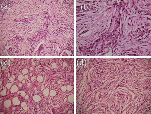
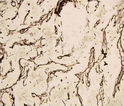
Figure 3. Photomicrograph of patient 3 showing reticulin positivity in stroma on immunohistochemistry (H&E×100)