Figures & data
Figure 1. T2 weighted MRI images obtained perpendicular to the long axis of the rectal tumour (a) before and (b) after short course radiotherapy showing reduction in tumour width post radiotherapy. In this case, tumour width reduced by 52% following short course radiotherapy. The images are obtained are at exactly matched levels as shown by the position of an adjacent lymph node (curved arrow) and vessels (arrow). The markers show how multiple radial measurements of tumour thickness were obtained to calculate the overall width measurement as a sum of the longest diameters (sum LD) according to RECIST criteria. The position of the mesorectal fascia is shown (arrow heads).
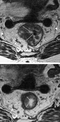
Figure 2. Sagittal T2 weighted MRI through rectal tumour (a) before and (b) after short course radiotherapy showing reduction in tumour length post treatment. In this case, tumour length reduced by 33% following short course radiotherapy.
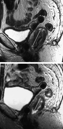
Figure 3. Scatter charts showing reduction in tumour (a) length and (b) width for each individual patient as shown by change in tumour (a) length and (b) width (y-axis) plotted against pre treatment (a) length and (b) width (x-axis). All points above the x axis are those which reduced in size. Those on or below the x axis either did not change or demonstrated a measurable increase in size.
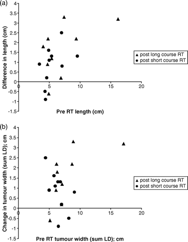
Figure 4. Oblique axial T2 weighted MRI through rectal tumour (a) before and (b) after short course radiotherapy. (a) Tumour invasion was thought to be just through the muscle layer (stage T3) with loss of normal low signal muscle wall (arrows). (b) Following short course radiotherapy the low signal inner and outer muscle layers (arrows) are now visible (stage T2 on MR but T1 on histology), suggesting downstaging.
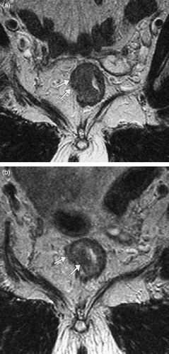
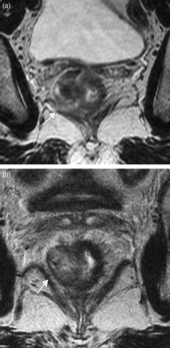
Figure 5. Oblique axial T2 weighted MRI through a low rectal tumour in a different patient (a) before and (b) after short course radiotherapy. (a) Tumour is seen to abut, possibly invade the right levator plate initially (arrow) with an ‘at risk’ crm. (b) Following short course radiotherapy there is a clear fat plane between the tumour (with intact muscle layer) and the levator muscle (arrow) and the crm is preserved.