3,005
Views
70
CrossRef citations to date
0
Altmetric
ORIGINAL ARTICLE

Accuracy of MRI for estimating residual tumor size after neoadjuvant chemotherapy in locally advanced breast cancer: Relation to response patterns on MRI

, , , , , , , , , , , , , & show all
Pages 996-1003 | Received 19 Oct 2007, Published online: 08 Jul 2009

Figures & data

Table I.  Patient characteristics before treatment.

Figure 1.  MRI before (A) and after (B) chemotherapy in a case accurately predicting residual disease. The tumor mass, measuring 1.7×2.0 cm before chemotherapy (A), disappeared completely on MRI after chemotherapy (B).
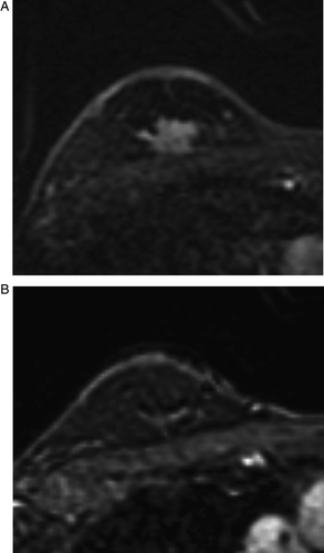
Figure 1.  MRI before (A) and after (B) chemotherapy in a case accurately predicting residual disease. The tumor mass, measuring 1.7×2.0 cm before chemotherapy (A), disappeared completely on MRI after chemotherapy (B).

Table II.  Tumor size expressed as the product of the two largest perpendicular diameters.

Figure 2.  Scatter plot of the largest tumor diameters measured by pathology versus those measured by MRI after chemotherapy.
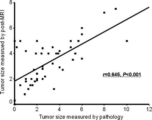
Figure 2.  Scatter plot of the largest tumor diameters measured by pathology versus those measured by MRI after chemotherapy.

Figure 3.  MRI before (A) and after (B) chemotherapy in a case showing a shrinkage pattern of the tumor response. The mass, measuring as 5.0×3.2 cm before chemotherapy (A), decreased to 1.5×1.5 cm and showed a shrinkage pattern of MRI after chemotherapy (B). The size of the residual tumor according to the pathologic assessment agreed with that determined by MRI, 1.4 cm maximum.
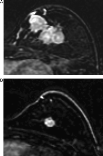
Figure 3.  MRI before (A) and after (B) chemotherapy in a case showing a shrinkage pattern of the tumor response. The mass, measuring as 5.0×3.2 cm before chemotherapy (A), decreased to 1.5×1.5 cm and showed a shrinkage pattern of MRI after chemotherapy (B). The size of the residual tumor according to the pathologic assessment agreed with that determined by MRI, 1.4 cm maximum.

Figure 4.  MRI before (A) and after (B) chemotherapy in a case showing nest pattern of the tumor response. The mass, measuring as 5.3×5.0 cm before chemotherapy (A), decreased slightly to 4.0×3.7 cm and showed a nest pattern on MRI after chemotherapy (B). A single inclusive measurement was given owing to multifocal enhancement after chemotherapy. The pathologic finding was a 1.8 cm, invasive ductal cancer with extensive intraductal components in a diffuse, unmeasurable, necrotic grayish area. MRI overestimated the size of the residual invasive tumor because it could not differentiate invasive from intraductal cancers
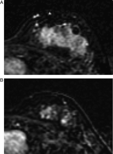
Figure 4.  MRI before (A) and after (B) chemotherapy in a case showing nest pattern of the tumor response. The mass, measuring as 5.3×5.0 cm before chemotherapy (A), decreased slightly to 4.0×3.7 cm and showed a nest pattern on MRI after chemotherapy (B). A single inclusive measurement was given owing to multifocal enhancement after chemotherapy. The pathologic finding was a 1.8 cm, invasive ductal cancer with extensive intraductal components in a diffuse, unmeasurable, necrotic grayish area. MRI overestimated the size of the residual invasive tumor because it could not differentiate invasive from intraductal cancers

Figure 5.  MRI before (A) and after (B) chemotherapy in a case showing a rim pattern of the tumor response. The mass, measuring as 1.8×2.0 cm before chemotherapy (A), decreased to 1.0×1.2 cm and showed a rim pattern on MRI after chemotherapy (B). The pathologic finding was IDC and measured 3.5 cm.
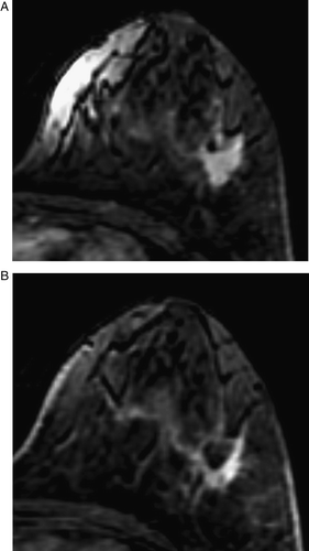
Figure 5.  MRI before (A) and after (B) chemotherapy in a case showing a rim pattern of the tumor response. The mass, measuring as 1.8×2.0 cm before chemotherapy (A), decreased to 1.0×1.2 cm and showed a rim pattern on MRI after chemotherapy (B). The pathologic finding was IDC and measured 3.5 cm.

Table III.  Agreement between the tumor sizes by MRI and by pathology, according to the tumor pattern.

Table IV.  Tumor patterns on MRI and the pathologic findings in the 14 over or underestimated cases.

Reprints and Corporate Permissions

Please note: Selecting permissions does not provide access to the full text of the article, please see our help page How do I view content?

To request a reprint or corporate permissions for this article, please click on the relevant link below:

Academic Permissions

Please note: Selecting permissions does not provide access to the full text of the article, please see our help page How do I view content?

Obtain permissions instantly via Rightslink by clicking on the button below:

If you are unable to obtain permissions via Rightslink, please complete and submit this Permissions form. For more information, please visit our Permissions help page.