3,784
Views
50
CrossRef citations to date
0
Altmetric
Original Article

Hodgkin lymphoma-related vanishing bile duct syndrome and idiopathic cholestasis: Statistical analysis of all published cases and literature review

, , , , , & show all
Pages 962-970 | Received 25 May 2007, Published online: 08 Jul 2009

Figures & data

Figure 1.  Axial CT scan of the chest with IV contrast showing a large anterior mediastinal soft-tissue mass measuring approximately 6.5 x 4.5 cm (depicted by white arrow).

Figure 1.  Axial CT scan of the chest with IV contrast showing a large anterior mediastinal soft-tissue mass measuring approximately 6.5 x 4.5 cm (depicted by white arrow).

Figure 2.  Liver biopsy, photographs taken of Masson trichrome-stained slides at 100× original magnification. 2a) Bile duct injury: this photograph shows a complete portal triad demonstrating bile duct injury. The bile duct (arrow) shows distortion of the duct with loss of nuclei. In this injured duct, only two nuclei remain. The hepatic artery branch is just above the duct cross section. 2b) Bile duct loss: this photograph shows an artery that is similar in appearance to the artery in a (it is the structure in the middle of the portal tract in both cases). But, in contrast to the other photo, there is no bile duct cross-section in this portal tract, representing destruction of the bile duct.

Figure 2.  Liver biopsy, photographs taken of Masson trichrome-stained slides at 100× original magnification. 2a) Bile duct injury: this photograph shows a complete portal triad demonstrating bile duct injury. The bile duct (arrow) shows distortion of the duct with loss of nuclei. In this injured duct, only two nuclei remain. The hepatic artery branch is just above the duct cross section. 2b) Bile duct loss: this photograph shows an artery that is similar in appearance to the artery in Figure 2a (it is the structure in the middle of the portal tract in both cases). But, in contrast to the other photo, there is no bile duct cross-section in this portal tract, representing destruction of the bile duct.

Table I.  Summary of Reported Literature.

Figure 3.  Kaplan-Meier survival curves for overall survival (OS) and liver failure-free survival (LFFS).

Figure 3.  Kaplan-Meier survival curves for overall survival (OS) and liver failure-free survival (LFFS).

Figure 4.  Kaplan-Meier liver failure-free survival curves correlated to stage of Hodgkin lymphoma (4a), radiation treatment (4b), and HL response (4c). Abbreviations: LFFS = liver failure-free survival, HL = Hodgkin lymphoma, CR = complete response. P-values calculated using the log-rank test.

Figure 4.  Kaplan-Meier liver failure-free survival curves correlated to stage of Hodgkin lymphoma (4a), radiation treatment (4b), and HL response (4c). Abbreviations: LFFS = liver failure-free survival, HL = Hodgkin lymphoma, CR = complete response. P-values calculated using the log-rank test.

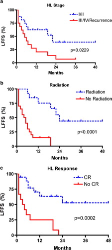
Table II.  Univariate Analysis for Liver Failure–Free Survival.

Reprints and Corporate Permissions

Please note: Selecting permissions does not provide access to the full text of the article, please see our help page How do I view content?

To request a reprint or corporate permissions for this article, please click on the relevant link below:

Academic Permissions

Please note: Selecting permissions does not provide access to the full text of the article, please see our help page How do I view content?

Obtain permissions instantly via Rightslink by clicking on the button below:

If you are unable to obtain permissions via Rightslink, please complete and submit this Permissions form. For more information, please visit our Permissions help page.