Figures & data
Figure 1. Saggital scout image using standard high resolution protocol: 3 mm slice interval, 16–18 cm FOV, 4–6 NSA, 256×256 matrix, TR >3,000, TE 80–100, ETL 16. In plane resolution 0.6×0.6 mm.
Figure 2. Detailed anatomy of bowel wall and tumour seen on high-resolution MRI.
Figure 3. High resolution T2-weighted axial MRI through rectal tumour showing the fascia propria of the mesorectum (red arrowheads) which corresponds to the surgical circumferential resection margin.
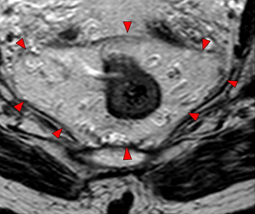
Figure 4. Standardised proforma for reporting of rectal MRI.
Figure 5. CRM is safe if distance of tumour to mesorectal fascia is >1 mm.
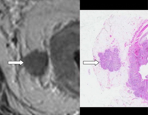
Figure 6. A tumour nodule is seen apparently extending laterally from the right side of the primary tumour. There is no histological evidence that this nodule is associated with any vascular structure on this slide. Serial ascending axial MRI slices through the same tumour suggest that the nodule lies within a tubular structure running parallel to the bowel wall, and the upper-most image shows signal void indicating that this is likely to be a vein.
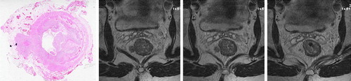
Table I. Summary of MRI-EMVI scoring system.
Figure 7. The presence of EMVI is associated with a significant reduction in relapse-free survival whether it is identified pre-operatively (upper graph) or histologically (lower graph). (Fig 7: Reproduced with permission. N. J. Smith, Y. Barbachano, A. R. Norman, R. I. Swift, A. M. Abulafi, G. Brown. Permission is granted by John Wiley & Sons Ltd on behalf of the BJSS Ltd Copyright # 2007 British Journal of Surgery Society Ltd).
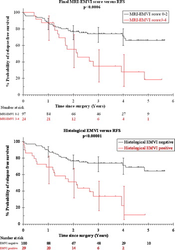
Figure 8. Extramural venous invasion lying close to the mesorectal resection margin. The image on the right shows the same level after chemoradiotherapy with some downstaging evident.
Figure 9. MRI-directed pre-operative treatment stratification as used in the EXPERT study.