Figures & data
Figure 1. Selection of regions of interests (ROIs). The tumor was transplanted on the right rear foot of 10–14 weeks old female CDF1 mice. The animals were restrained in a plastic jig. The right foot was exposed and secured with tape, and the tail was put through a hole in the rear of the jig, helping to secure the mouse in a fixed position. The tumor was positioned adjacent to a radiofrequency surface-coil Citation[13]. DWI, PWI and T2 weighted imaging was acquired with a coronal slice, which has been stated in the text. This means that the slice orientation was mostly parallel to the surface of the skin. The area in images representing tumour tissue was divided into three regions of interests: 1) whole tumour segment, a ROI covering the whole tumour area, 2) high perfusion segments, and 3) low perfusion segments. The tumour areas with the lowest blood perfusion were primarily located in the central parts of the tumour, whereas higher levels of blood perfusion were found in the periphery close to the surrounding healthy cells.
![Figure 1. Selection of regions of interests (ROIs). The tumor was transplanted on the right rear foot of 10–14 weeks old female CDF1 mice. The animals were restrained in a plastic jig. The right foot was exposed and secured with tape, and the tail was put through a hole in the rear of the jig, helping to secure the mouse in a fixed position. The tumor was positioned adjacent to a radiofrequency surface-coil Citation[13]. DWI, PWI and T2 weighted imaging was acquired with a coronal slice, which has been stated in the text. This means that the slice orientation was mostly parallel to the surface of the skin. The area in images representing tumour tissue was divided into three regions of interests: 1) whole tumour segment, a ROI covering the whole tumour area, 2) high perfusion segments, and 3) low perfusion segments. The tumour areas with the lowest blood perfusion were primarily located in the central parts of the tumour, whereas higher levels of blood perfusion were found in the periphery close to the surrounding healthy cells.](/cms/asset/6e796d8d-03ca-4a1d-98d6-a77bdc4932f9/ionc_a_276967_f0001_b.gif)
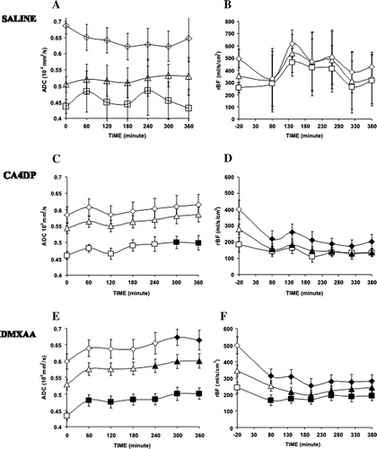
Figure 2. Time dependent effects of saline (0.4 ml ip), CA4DP (250 mg/kg ip) and DMXAA (20 mg/kg ip) on ADC (2A, 2C, 2E) and relative blood flow (rBF) (2B + 2D + 2F). Results are shown following treatment with saline (2A, 2B), CA4DP (2C, 2D) or DMXAA (2E, 2F) for high-perfused (⋄), low-perfused (□) and whole tumour areas (Δ). Values that are significantly different compared to baseline values are filled with black color. Data are shown as mean±sem.