Figures & data
Figure 1. Sagital T1-weighted MRI of the thoracic spine showing marrow infiltration, endplate destruction, and paravertebral mass at T5 and T6 level. Note involvement of anterior longitudinal ligament.
Figure 2. Axial view.
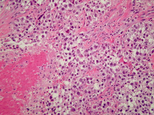
Figure 3. Paraspinal tumor. The clear cell sarcoma is comprised of polygonal cells with abundant clear to pale cytoplasm bordered by thin fibrous septae. A small amount of associated necrosis is seen in the lower left hand corner. (H & E section, 200X)