3,022
Views
76
CrossRef citations to date
0
Altmetric
Original Article

Morphology, metabolism, microcirculation, and strength of skeletal muscles in cancer-related cachexia

, , , , , , , , & show all
Pages 116-124 | Received 03 Dec 2007, Published online: 08 Jul 2009

Figures & data

Figure 1.  Magnetic resonance T1-weighted imaging of the right thigh in a 54-year old man with cachexia due to pancreatic cancer (weight 58 kg) (A) and the corresponding right thigh at the same level of the matched healthy volunteer (55 years, weight 107 kg) (B). In A), determination of muscle cross-sectional area (CSA) of the right quadriceps femoris is illustrated (CSA in patient with cachexia vs. volunteer, 51.3 vs. 85.9 cm2). In B), the quantification of the ratio of signal intensities of muscle and subcutaneous fat tissue using a region-of-interest analysis is illustrated. The corresponding 1H MR spectrum (C) from the right vastus lateralis muscle and the 31P MR spectrum of the same region (D) of the patient with cachexia illustrate the metabolites that were quantified. Peak assignments in C): Cr, (phospho-)creatine; TMA peak, trimethyl-ammonium-containing compounds among which is choline; IMCL, intramyocellular lipids; EMCL, extramyocellular lipids. Peak assignments in D): Phosphocreatine (PCr), inorganic phosphate (Pi), phosphodiester (PDE), and the three resonances (α, β, γ) of adenosine 5′-triphosphate (ATP); intensity ratio β-ATP/PCr in patient with cachexia vs. volunteer, 0.27 vs. 0.27. Only the signals printed in bold in C) and D) were quantified. The corresponding transverse power Doppler images after bolus injection of 10 ml Levovist® in 1.5 cm depth (focus area) show the initial increase (E) and maximum plateau (F) of microbubbles’ replenishment within the right vastus lateralis muscle of the patient with cachexia. Microcirculation and concentration of lipid and energy metabolites in vivo were comparable in muscles of volunteers and cachectic patients at rest.

Figure 1.  Magnetic resonance T1-weighted imaging of the right thigh in a 54-year old man with cachexia due to pancreatic cancer (weight 58 kg) (A) and the corresponding right thigh at the same level of the matched healthy volunteer (55 years, weight 107 kg) (B). In A), determination of muscle cross-sectional area (CSA) of the right quadriceps femoris is illustrated (CSA in patient with cachexia vs. volunteer, 51.3 vs. 85.9 cm2). In B), the quantification of the ratio of signal intensities of muscle and subcutaneous fat tissue using a region-of-interest analysis is illustrated. The corresponding 1H MR spectrum (C) from the right vastus lateralis muscle and the 31P MR spectrum of the same region (D) of the patient with cachexia illustrate the metabolites that were quantified. Peak assignments in C): Cr, (phospho-)creatine; TMA peak, trimethyl-ammonium-containing compounds among which is choline; IMCL, intramyocellular lipids; EMCL, extramyocellular lipids. Peak assignments in D): Phosphocreatine (PCr), inorganic phosphate (Pi), phosphodiester (PDE), and the three resonances (α, β, γ) of adenosine 5′-triphosphate (ATP); intensity ratio β-ATP/PCr in patient with cachexia vs. volunteer, 0.27 vs. 0.27. Only the signals printed in bold in C) and D) were quantified. The corresponding transverse power Doppler images after bolus injection of 10 ml Levovist® in 1.5 cm depth (focus area) show the initial increase (E) and maximum plateau (F) of microbubbles’ replenishment within the right vastus lateralis muscle of the patient with cachexia. Microcirculation and concentration of lipid and energy metabolites in vivo were comparable in muscles of volunteers and cachectic patients at rest.

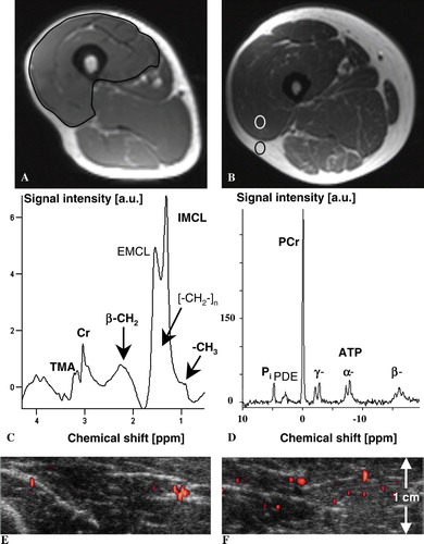
Table I.  Overview of data from cancer-related cachectic patients and healthy volunteers.

Reprints and Corporate Permissions

Please note: Selecting permissions does not provide access to the full text of the article, please see our help page How do I view content?

To request a reprint or corporate permissions for this article, please click on the relevant link below:

Academic Permissions

Please note: Selecting permissions does not provide access to the full text of the article, please see our help page How do I view content?

Obtain permissions instantly via Rightslink by clicking on the button below:

If you are unable to obtain permissions via Rightslink, please complete and submit this Permissions form. For more information, please visit our Permissions help page.