Figures & data
Table I. Sequences of the primers used in amplifying SSTR1-5 cDNA and the length of each fragment amplified.
Figure 1. The expression of SSTRs in capan-2 and A549 cells. A: The RNA expressions of all five SSTRs were analyzed by RT-PCR. SSTR1 (lane 1), SSTR2 (lane 2), SSTR4 (lane 3) and SSTR5 (lane 4) were expressed in capan-2 cells while SSTR1 (lane 5) and SSTR3 (lane 6) were expressed in A549 cells. DNA ladders were loaded in lane M.B: The protein expression of SSTR2 was detected in capan-2 (3) and A549 cells (4) transfected with Adv-SSTR2, shown by western blotting with anti-SSTR2. No SSTR2 proteins were detected in control capan-2 (1) cells or A549 (2) cells transfected with Adv-LacZ. The cellular β-actin was shown as an internal control.
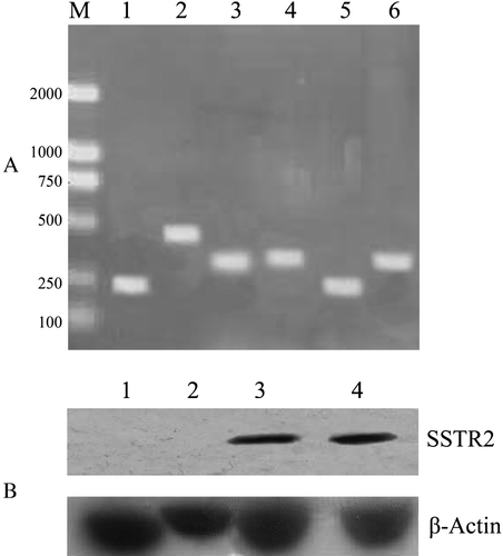
Figure 2. The efficacy of transfection of the constructed adenovirus vector in cultured cells. The transfection efficacy of the adenovirus vectors was demonstrated with Adv-LacZ using the β-galactosidase staining. A: untransfected capan-2 cells. B: untransfected A549 cells. C: Adv-LacZ transfected capan-2 cells. D: Adv-LacZ transfected A549 cells. The cells showed positive β-galactosidase staining were visualized under microscope (x 200) and nearly 100% transfection was shown when MOI was up to 100. Untransfected cells showed negative β-galactosidase staining.
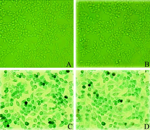
Figure 3. The transfection efficacy of intratumoral injection of the adenovirus. The cells that were successfully transfected by intratumoral injection of Adv-LacZ (C) or Adv-SSTR2 (D) in capan-2 xenografts were shown by β-galactosidase staining and histrochemistry, respectively. The results were observed under light microscope (x 400). A: untransfected conrol of capan-2 xenograft, stained for β-galactosidase; B: untransfected control of capan-2 xenograft stained with anti-SSTR2 and HRP-conjugated secondary antibody; C: capan-2 xenograft transfected with Adv-LacZ, stained for β-galactosidase; D: capan-2 xenograft transfected with Adv-SSTR2 was stained with anti-SSTR2 and HRP-conjugated secondary antibody.
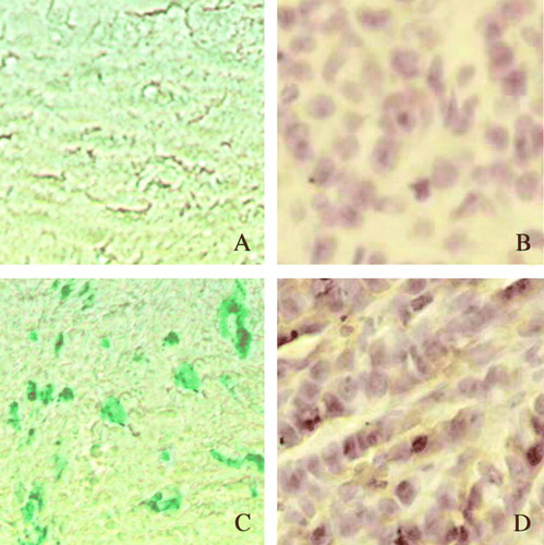
Figure 4. Cancer cells overexpressing exogenous hSSTR2 failed in tumor formation. Capan-2 cells and A549 cells overexpressing exogenous human SSTR2 failed in tumor formation in all our experimental nude mice. MOI=100 was used in in vitro transfection of cultured cells. A: No discernable tumors were observed with the injected capan-2 cells transfected with Adv-SSTR2 (indicated with white arrows), whereas successful tumor formations were observed with the injected capan-2 cells transfected with Adv-LacZ (indicated with red arrows). B: A: No discernable tumors were observed with the injected A549 cells transfected with Adv-SSTR2 (indicated with white arrows), whereas successful tumor formations were observed with the injected A549 cells transfected with Adv-LacZ (indicated with red arrows).
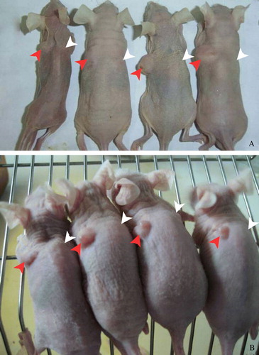
Figure 5. Overexpression of hSSTR2 inhibited the growth of human pancreatic tumors. The intratumoral injection of the Adv-SSTR2 in the capan-2 xenografts resulted in marked inhibition of the tumor growth. A: Adv-SSTR2 were injected into the capan-2 xenografts (indicated with white arrows) while Adv-LacZ was injected into the controls (indicated with red arrows). B: The tumor tissues, Adv-LacZ transfected (upper panel) and Adv-SSTR2 transfected (lower panel), were excised and measured four weeks after the last injection. C: The overexpressed SSTR2 was detected in capan-2 xenografts transfected with Adv-SSTR2 (lane 2) but not in Adv-LacZ transfected controls (lane 1), shown by Western Blot assay (lane 2) while it was using anti-SSTR2 and HRP-conjugated secondary antibody. The endogenous β-actin was used as a control.
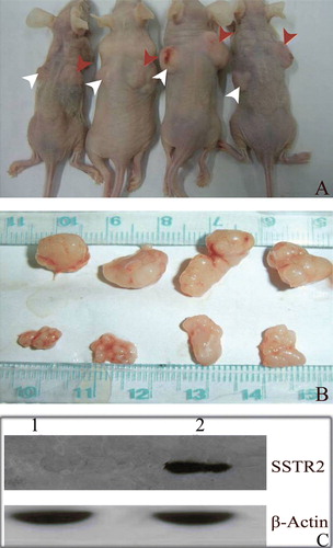
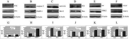
Figure 6. Overexpression of SSTR2 affected multiple components in different pathways. The influences of SSTR2 overexpression on components in a number of different pathways were analyzed in Adv-SSTR2 transfected capan-2 xenografts using western blotting. The control capan-2 xenografts were transfected with Adv-LacZ and the expression of β-Actin was used as a sample loading control. The overexpression of SSTR2 was confirmed in the Adv-SSTR2 transfected capan-2 xenograft by western (Lane 2) while was not observed in the Adv-LacZ transfected control (Lane 1), as shown in . The expressions of p16, p21, caspase-3, PARP-1, p53, Bax, survivin, Bcl-2, ras, ERK2, VEGF and TIMP-2 in both Adv-SSTR2 transfected capan-2 xenografts and Adv-LacZ transfected controls were monitored by western blot assay (A, B, C, D, E and F). The results of the western blot assay were analyzed by AlphaEase FC StandAlone and the cellular levels of the above molecules were compared between the Adv-SSTR2 transfected tumors and the Adv-LacZ transfected controls (G, H, I, J, K and L).