Figures & data
Figure 1. 1A. A lymphoscintigram with four sentinel nodes of an 11-year-old girl with a melanoma in the middle of the left cheek. The primary lesion (Breslow depth 3.8 mm, Clark level IV) had originated in a benign Spitz nevus. 1B. A blue-stained sentinel node (II sn) found in the parotid gland. At the same time of SNB, the biopsy scar of the primary lesion was excised with 1.5 cm lateral margins and the wound was closed directly. 1C. Histopathological analysis (hematoxylin and eosin staining) revealed a subcapsular micrometastasis with minimal tumour burden. It was the only metastatic sentinel node. In a second stage operation, superficial parotidectomy and selective neck dissection of levels II–III were performed and no additional metastatic nodes were detected. 1D. Aesthetic result two years after the operation. The patient has remained disease-free after a follow-up of 27 months.
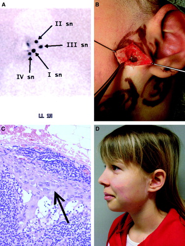
Table I. Distribution of sentinel nodes according to the location of the primary lesion
Table II. Comparison between clinical and histopathological characteristics of SNB patients and control patients
Table III. Distribution of metastatic lymph node basins according to the location of primary lesion
Figure 2. Melanoma-specific overall survival of 25 SNB patients, 29 ELND patients and 92 observational patients.
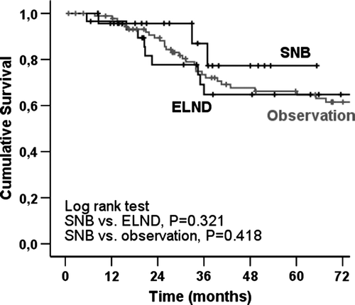
Figure 3. Melanoma-specific overall survival of 77 female and 69 male patients with H&N melanoma.
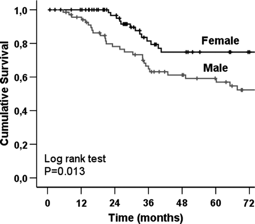
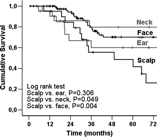
Figure 4. Melanoma-specific overall survival of 146 H&N melanoma patients according to the anatomic site of the primary lesion.