Figures & data
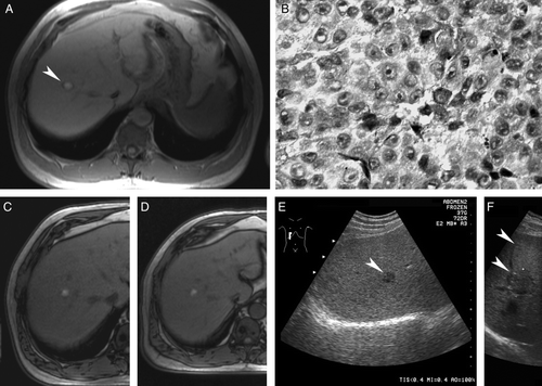
Figure 1. Liver metastasis from uveal melanoma. Magnetic resonance imaging (MRI) shows a solitary 20 mm target lesion (arrowhead) in the right lobe of the liver (A). Core needle biopsy confirms metastatic melanoma (B; hematoxylin-eosin). MRI documents partial response after the 3rd (C) cycle, maintained after the 9th cycle and subsequent interferon monotherapy (D). Same lesion seen with ultrasonography before (E; arrowhead) and after (F) adjuvant thermoablation (small crosses) with the ablated needle track (arrowheads).