Figures & data
Figure 1. Haggit's sub-classification of polyp-cancers. The stage is related to the extent of penetration into the stalk of the polyp.
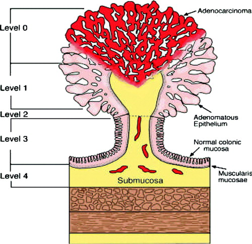
Figure 2. Sub-classification of T1 cancers based upon depth of invasion into the submucosal layer.
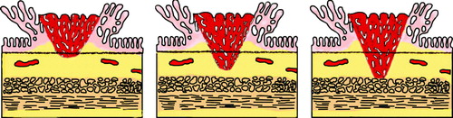
Table I. Elements to be included in the pathological report on T1 rectal adenocarcinoma.
Table II. Accuracy of T and N staging by endoscopic ultrasound in patients with rectal cancer.
Figure 3. The conventional 10 MHz B mode ultrasound picture is shown to the right and the sonoelastographic picture to the left. Note the colour scale indicating the strain as hard (blue to soft (red). The read and green colour of the tumour indicates that it is softer than the surrounding normal bowel wall, which is blue.
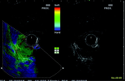
Figure 4. Endoscopic submucosal dissection of adenomas and polypoid low risk T1 cancers of Haggits stage 1 or 2.
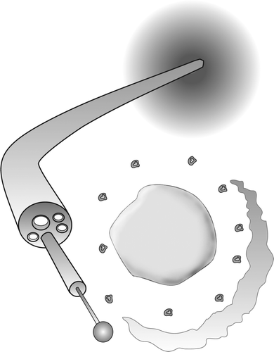
Figure 5. The TEM equipment for transanal stereomicroscopic dissection of low risk T1 cancers.
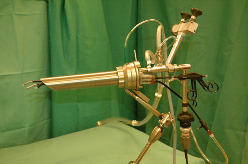
Table III. Reported local recurrence rates after TEM-resection of T1 cancer. Non-comparative case-series including at least 20 patients.
Table IV. Factors indicating a good prognosis in T1 cancer.