Figures & data
Figure 1. Images of left thigh and left calf, over time. (A) February 2015, before electrochemotherapy. Cutaneous metastases extended from the left side of the abdomen to the left calf, with bleeding lesions. (B) March 2015, the day after treatment with electrochemotherapy. The treated areas include the front and back of her thigh, 20 × 25 cm and 30 × 13 cm, respectively, and on the back of her calf 20 × 10 cm. The areas are marked with pen and appear erythematous. The bleeding has stopped and the metastases appear pale. (C) May 2015, two months after treatment with electrochemotherapy, leveling of the cutaneous metastases in the treated area, the lesions appear more pale and as scars, from where exophytic lesions have fallen off, are visible. (D) July 2016, 12 months after retreatment with calcium and electroporation and 16 months after the first electrochemotherapy, complete leveling of all cutaneous metastases and appearance of vitiligo in both treated and untreated area. Biopsies from pigmented lesions were without malignancy (see supplementary Figure 1).
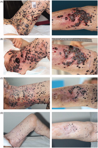
Figure 2. July 2016. Illustration of widespread vitiligo in treated and untreated areas. (A) Vitiligo in distant untreated area. (B–D) Vitiligo extends beyond the actual treated area.
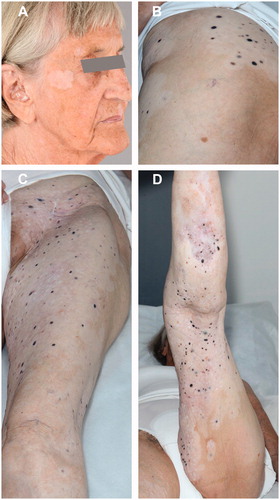
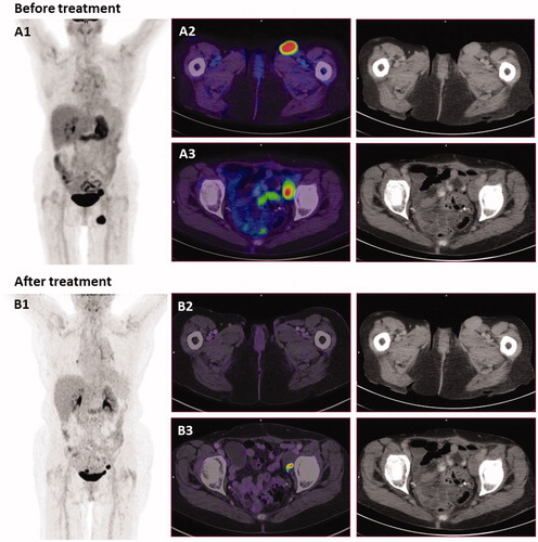
Figure 3. PET-CT scans and CT-scans before and after treatment. (A) September 2014, before treatment with electrochemotherapy. (A1) MIP image (maximum intensity projection), showing the two metastases. (A2) PET/CT scan shows an enlarged lymph node in the left inguinal region measuring 2.7 cm. (A3) An enlarged necrotic lymph node in the left side of pelvis measuring 2.4 cm. (B) July 2016, 16 months after electrochemotherapy and 12 months after retreatment with calcium electroporation and electrochemotherapy. (B1) MIP image shows disappearance of lymph node in the left inguinal region. (B2) PET/CT scan shows disappearance of lymph node in left inguinal region. (B3) Lymph node in left pelvis is almost completely necrotic, and cannot be measured. This inguinal node was subsequently biopsied and there was no sign of malignancy. Of note, a new PET/CT-scanner was employed for the second scan, thus direct comparison of the SUV values is not strictly possible.