Figures & data
Figure 1. Immunohistochemical staining for PHPT1. A: An E14.5 mouse embryo sagittal section shows PHPT1 signal in the epithelium of the choroid plexus in the fourth ventricle of brain (arrow). B: Adjacent section of A, using preimmune serum as a negative control. C: The same expression pattern of PHPT1 was found in the epithelium layer of the choroid plexus in an adult mouse brain (arrow). Arrowheads point at the ependymal cells in the ventricle. D: Purkinje cell (arrow) in the cerebellum expressing PHPT1. E: PHPT1 expression in the E14.5 embryonic heart muscle. F: PHPT1 expression (arrow) in the epithelium layer of the developing gut of an E14.5 sagittal section. Amplifications were 100× for A, B, and E, 400× for C and D, and 25× for F.
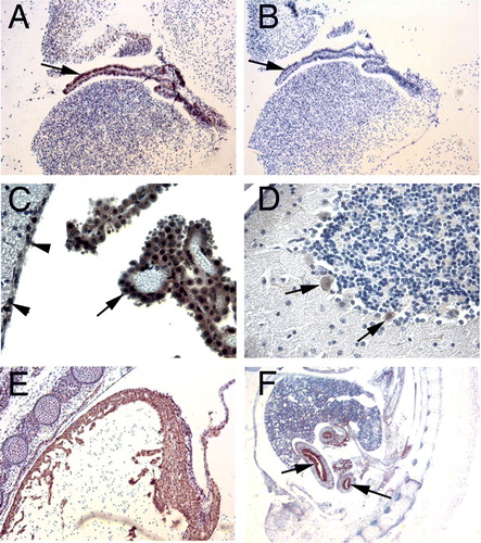
Figure 2. Immunohistochemical staining for PHPT1. A: A section of adult mouse kidney shows PHPT1 expression in the distal convoluted tubules (arrowhead) but not in the glomeruli (arrow) and a weak expression in the proximal convoluted tubule. B: PHPT1 is expressed in the Henle's loops of adult kidney. C and D: Sections of seminiferous tubule of adult mouse testis. Arrows in C point to the spermatogonium of seminiferous tubule. D: An absorption test shows that PHPT1 signals were abolished in the mouse testis. E: PHPT1 is expressed in the epithelium of interlobular duct of pancreas (arrow). F: PHPT1 is expressed in the epithelium of bronchiole (arrow). Arrowheads point at macrophages in alveoli. Amplifications were 400× for A–F.
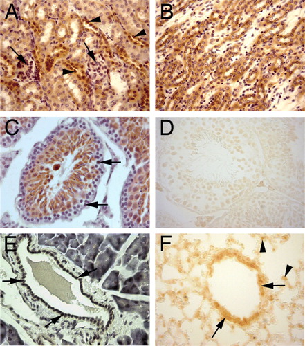
Table I. Protein expression of PHPT1 in mouse and human tissues.