550
Views
11
CrossRef citations to date
0
Altmetric
Case Report

Langerhans cell histiocytosis of the sternum

, MD, , , , &
Pages 121-125 | Received 05 Dec 2008, Published online: 12 Jul 2009

Figures & data

Figure 1.  A lateral radiograph of the sternum showed an ill-defined osteolytic lesion in the sternum. Periosteal reactions around the lesion were also observed.

Figure 1.  A lateral radiograph of the sternum showed an ill-defined osteolytic lesion in the sternum. Periosteal reactions around the lesion were also observed.

Figure 2.  Computed tomography (CT) showed an osteolytic lesion that dissolved both anterior and posterior cortex of the body of the sternum.

Figure 2.  Computed tomography (CT) showed an osteolytic lesion that dissolved both anterior and posterior cortex of the body of the sternum.

Figure 3.  Magnetic resonance imaging (MRI). The signal intensity of the tumor was low in the sternum on T1-weighted images (A) and high on T2-weighted images (B).

Figure 3.  Magnetic resonance imaging (MRI). The signal intensity of the tumor was low in the sternum on T1-weighted images (A) and high on T2-weighted images (B).

Figure 4.  Technetium bone scintigraphy reveals increased uptakes in the sternum, the greater trochanter of the right femur, and the right distal tibia.

Figure 4.  Technetium bone scintigraphy reveals increased uptakes in the sternum, the greater trochanter of the right femur, and the right distal tibia.

Figure 5.  Radiographs show small and round osteolytic lesions (arrows) in the greater trochanter of the right femur (A) and the distal tibia (B).

Figure 5.  Radiographs show small and round osteolytic lesions (arrows) in the greater trochanter of the right femur (A) and the distal tibia (B).

Figure 6.  Histopathologic findings. Histopathologic examination reveals a proliferation of histiocytes with an infiltration of eosinophils. These histocytes were positive for S-100 (B), and for CD-1a (C). Original magnification ×400.

Figure 6.  Histopathologic findings. Histopathologic examination reveals a proliferation of histiocytes with an infiltration of eosinophils. These histocytes were positive for S-100 (B), and for CD-1a (C). Original magnification ×400.

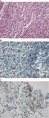
Table I.  Literature of sternal lesion of LCH.

Reprints and Corporate Permissions

Please note: Selecting permissions does not provide access to the full text of the article, please see our help page How do I view content?

To request a reprint or corporate permissions for this article, please click on the relevant link below:

Academic Permissions

Please note: Selecting permissions does not provide access to the full text of the article, please see our help page How do I view content?

Obtain permissions instantly via Rightslink by clicking on the button below:

If you are unable to obtain permissions via Rightslink, please complete and submit this Permissions form. For more information, please visit our Permissions help page.